


X-ray structure of a RNase S variant in complex with an artificial peptide
Gao, J., Genz, M., Hofman, F., Hiller, S., Strater, N., Seebeck, F.To be published.
Experimental Data Snapshot
Starting Model: experimental
View more details
wwPDB Validation 3D Report Full Report

Entity ID: 1 | |||||
|---|---|---|---|---|---|
| Molecule | Chains | Sequence Length | Organism | Details | Image |
| Ribonuclease pancreatic | 15 | Bos taurus | Mutation(s): 2 EC: 3.1.27.5 (PDB Primary Data), 4.6.1.18 (UniProt) | 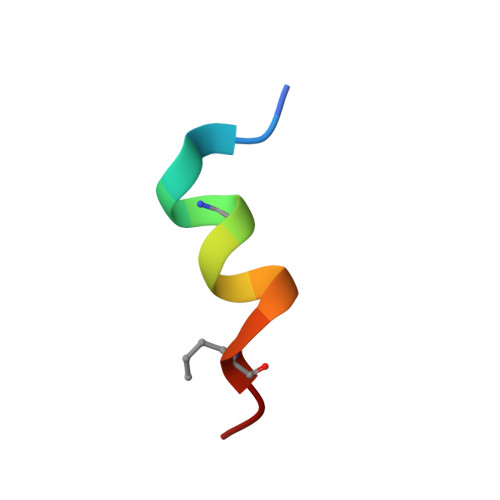 | |
UniProt | |||||
Entity Groups | |||||
| Sequence Clusters | 30% Identity50% Identity70% Identity90% Identity95% Identity100% Identity | ||||
| UniProt Group | P61823 | ||||
Sequence AnnotationsExpand | |||||
Reference Sequence | |||||
Entity ID: 2 | |||||
|---|---|---|---|---|---|
| Molecule | Chains | Sequence Length | Organism | Details | Image |
| Ribonuclease pancreatic | 104 | Bos taurus | Mutation(s): 0 EC: 3.1.27.5 (PDB Primary Data), 4.6.1.18 (UniProt) | 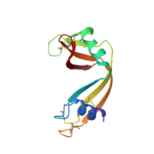 | |
UniProt | |||||
Entity Groups | |||||
| Sequence Clusters | 30% Identity50% Identity70% Identity90% Identity95% Identity100% Identity | ||||
| UniProt Group | P61823 | ||||
Sequence AnnotationsExpand | |||||
Reference Sequence | |||||
| Ligands 2 Unique | |||||
|---|---|---|---|---|---|
| ID | Chains | Name / Formula / InChI Key | 2D Diagram | 3D Interactions | |
| SO4 Download:Ideal Coordinates CCD File | C [auth A], G [auth B] | SULFATE ION O4 S QAOWNCQODCNURD-UHFFFAOYSA-L |  | ||
| CL Download:Ideal Coordinates CCD File | D [auth A] E [auth A] F [auth A] H [auth B] I [auth B] | CHLORIDE ION Cl VEXZGXHMUGYJMC-UHFFFAOYSA-M |  | ||
| Modified Residues 2 Unique | |||||
|---|---|---|---|---|---|
| ID | Chains | Type | Formula | 2D Diagram | Parent |
| DHA Query on DHA | A | PEPTIDE LINKING | C3 H5 N O2 |  | SER |
| NLE Query on NLE | A | L-PEPTIDE LINKING | C6 H13 N O2 |  | LEU |
| Length ( Å ) | Angle ( ˚ ) |
|---|---|
| a = 43.8 | α = 90 |
| b = 43.8 | β = 90 |
| c = 96.7 | γ = 120 |
| Software Name | Purpose |
|---|---|
| MAR345dtb | data collection |
| REFMAC | refinement |
| XDS | data reduction |
| XSCALE | data scaling |
| REFMAC | phasing |


