A pre-steady state and steady state kinetic analysis of the N-ribosyl hydrolase activity of hCD157.
Preugschat, F., Carter, L.H., Boros, E.E., Porter, D.J., Stewart, E.L., Shewchuk, L.M.(2014) Arch Biochem Biophys 564C: 156-163
- PubMed: 25250980 Search on PubMed
- DOI: https://doi.org/10.1016/j.abb.2014.09.008
- Primary Citation Related Structures:
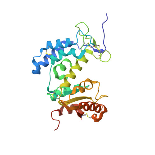
4OGW - PubMed Abstract:
hCD157 catalyzes the hydrolysis of nicotinamide riboside (NR) and nicotinic acid riboside (NAR). The release of nicotinamide or nicotinic acid from NR or NAR was confirmed by spectrophotometric, HPLC and NMR analyses. hCD157 is inactivated by a mechanism-based inhibitor, 2'-deoxy-2'-fluoro-nicotinamide arabinoside (fNR). Modification of the enzyme during the catalytic cycle by NR, NAR, or fNR increased the intrinsic protein fluorescence by approximately 50%. Pre-steady state and steady state data were used to derive a minimal kinetic scheme for the hydrolysis of NR. After initial complex formation a reversible step (360 and 30s(-1)) is followed by a slow irreversible step (0.1s(-1)) that defined the rate limiting step, or kcat. The calculated KMapp value for NR in the hydrolytic reaction is 6nM. The values of the kinetic constants suggest that one biological function of cell-surface hCD157 is to bind and slowly hydrolyze NR, possibly converting it to a ligand-activated receptor. Differences in substrate preference between hCD157 and hCD38 were rationalized through a comparison of the crystal structures of the two proteins. This comparison identified several residues in hCD157 (F108 and F173) that can potentially hinder the binding of dinucleotide substrates (NAD+).
- Muscle Metabolism Discovery Performance Unit, GlaxoSmithKline, 5 Moore Drive, 3.2094, Research Triangle Park, NC 27709, United States. Electronic address: Frank.X.Preugschat@gsk.com.
Organizational Affiliation: