4OAZ

BldD CTD-c-di-GMP complex


Experimental Data Snapshot

  • Method: X-RAY DIFFRACTION
  • Resolution: 2.25 Å
  • R-Value Free: 0.284 
  • R-Value Work: 0.236 
  • R-Value Observed: 0.243 

wwPDB Validation   3D Report Full Report


Ligand Structure Quality Assessment 


This is version 1.1 of the entry. See complete history


Literature

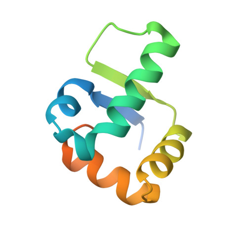
Tetrameric c-di-GMP mediates effective transcription factor dimerization to control Streptomyces development.

Tschowri, N.Schumacher, M.A.Schlimpert, S.Chinnam, N.B.Findlay, K.C.Brennan, R.G.Buttner, M.J.

(2014) Cell 158: 1136-1147

  • DOI: https://doi.org/10.1016/j.cell.2014.07.022
  • Primary Citation of Related Structures:  
    4OAY, 4OAZ, 4OB4, 5KHD

  • PubMed Abstract: 
  • The cyclic dinucleotide c-di-GMP is a signaling molecule with diverse functions in cellular physiology. Here, we report that c-di-GMP can assemble into a tetramer that mediates the effective dimerization of a transcription factor, BldD, which controls the progression of multicellular differentiation in sporulating actinomycete bacteria ...

    The cyclic dinucleotide c-di-GMP is a signaling molecule with diverse functions in cellular physiology. Here, we report that c-di-GMP can assemble into a tetramer that mediates the effective dimerization of a transcription factor, BldD, which controls the progression of multicellular differentiation in sporulating actinomycete bacteria. BldD represses expression of sporulation genes during vegetative growth in a manner that depends on c-di-GMP-mediated dimerization. Structural and biochemical analyses show that tetrameric c-di-GMP links two subunits of BldD through their C-terminal domains, which are otherwise separated by ~10 Å and thus cannot effect dimerization directly. Binding of the c-di-GMP tetramer by BldD is selective and requires a bipartite RXD-X8-RXXD signature. The findings indicate a unique mechanism of protein dimerization and the ability of nucleotide signaling molecules to assume alternative oligomeric states to effect different functions.


    Organizational Affiliation

    Department of Molecular Microbiology, John Innes Centre, Norwich Research Park, Norwich NR4 7UH, UK. Electronic address: mark.buttner@jic.ac.uk.


Macromolecules
Find similar proteins by:  (by identity cutoff)  |  3D Structure
Entity ID: 1
MoleculeChainsSequence LengthOrganismDetailsImage
Putative DNA-binding proteinA [auth R],
B [auth A]
92Streptomyces coelicolor A3(2)Mutation(s): 0 
Gene Names: SCO1489
UniProt
Find proteins for Q7AKQ8 (Streptomyces coelicolor (strain ATCC BAA-471 / A3(2) / M145))
Explore Q7AKQ8 
Go to UniProtKB:  Q7AKQ8
Entity Groups  
Sequence Clusters30% Identity50% Identity70% Identity90% Identity95% Identity100% Identity
UniProt GroupQ7AKQ8
Protein Feature View
Expand
  • Reference Sequence
Small Molecules
Ligands 1 Unique
IDChainsName / Formula / InChI Key2D Diagram3D Interactions
C2E
Query on C2E

Download Ideal Coordinates CCD File 
C [auth R],
D [auth R],
E [auth A],
F [auth A]
9,9'-[(2R,3R,3aS,5S,7aR,9R,10R,10aS,12S,14aR)-3,5,10,12-tetrahydroxy-5,12-dioxidooctahydro-2H,7H-difuro[3,2-d:3',2'-j][1,3,7,9,2,8]tetraoxadiphosphacyclododecine-2,9-diyl]bis(2-amino-1,9-dihydro-6H-purin-6-one)
C20 H24 N10 O14 P2
PKFDLKSEZWEFGL-MHARETSRSA-N
 Ligand Interaction
Experimental Data & Validation

Experimental Data

  • Method: X-RAY DIFFRACTION
  • Resolution: 2.25 Å
  • R-Value Free: 0.284 
  • R-Value Work: 0.236 
  • R-Value Observed: 0.243 
  • Space Group: C 2 2 21
Unit Cell:
Length ( Å )Angle ( ˚ )
a = 36.87α = 90
b = 95.53β = 90
c = 100.12γ = 90
Software Package:
Software NamePurpose
PHENIXrefinement

Structure Validation

View Full Validation Report


Ligand Structure Quality Assessment 


Entry History 

Deposition Data

Revision History  (Full details and data files)

  • Version 1.0: 2014-11-19
    Type: Initial release
  • Version 1.1: 2015-09-16
    Changes: Database references