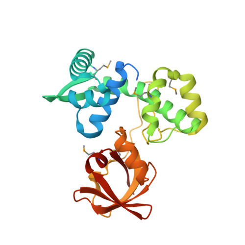
Structural analysis and insight into metal-ion activation of the iron-dependent regulator from Thermoplasma acidophilum.
Yeo, H.K., Park, Y.W., Lee, J.Y.(2014) Acta Crystallogr D Biol Crystallogr 70: 1281-1288
- PubMed: 24816097
- DOI: https://doi.org/10.1107/S1399004714004118
- Primary Citation of Related Structures:
4O5V, 4O6J - PubMed Abstract:
The iron-dependent regulator (IdeR) is a metal ion-activated transcriptional repressor that regulates the expression of genes encoding proteins involved in iron uptake to maintain metal-ion homeostasis. IdeR is a functional homologue of the diphtheria toxin repressor (DtxR), and both belong to the DtxR/MntR family of metalloregulators. The structure of Fe(2+)-bound IdeR (TA0872) from Themoplasma acidophilum was determined at 2.1 Å resolution by X-ray crystallography using single-wavelength anomalous diffraction. The presence of Fe(2+), which is the true biological activator of IdeR, in the metal-binding site was ascertained by the use of anomalous difference electron-density maps using diffraction data collected at the Fe absorption edge. Each DtxR/IdeR subunit contains two metal ion-binding sites separated by 9 Å, labelled the primary and ancillary sites, whereas the crystal structures of IdeR from T. acidophilum show a binuclear iron cluster separated by 3.2 Å, which is novel to T. acidophilum IdeR. The metal-binding site analogous to the primary site in DtxR was unoccupied, and the ancillary site was occupied by binuclear clustered ions. This difference suggests that T. acidophilum IdeR and its closely related homologues are regulated by a mechanism distinct from that of either DtxR or MntR. T. acidophilum IdeR was also shown to have a metal-dependent DNA-binding property by electrophoretic mobility shift assay.
- Department of Life Science, Dongguk University Seoul, 26 Pil-dong 3-ga, Jung-gu, Seoul 100-715, Republic of Korea.
Organizational Affiliation:


