4NYQ

In-vivo crystallisation (midguts of a viviparous cockroach) and structure at 1.2 A resolution of a glycosylated, lipid-binding, lipocalin-like protein


Experimental Data Snapshot

  • Method: X-RAY DIFFRACTION
  • Resolution: 1.20 Å
  • R-Value Free: 0.192 
  • R-Value Work: 0.143 
  • R-Value Observed: 0.146 

wwPDB Validation   3D Report Full Report


Ligand Structure Quality Assessment 


This is version 2.1 of the entry. See complete history


Literature

Structure of a heterogeneous, glycosylated, lipid-bound, in vivo-grown protein crystal at atomic resolution from the viviparous cockroach Diploptera punctata.

Banerjee, S.Coussens, N.P.Gallat, F.X.Sathyanarayanan, N.Srikanth, J.Yagi, K.J.Gray, J.S.Tobe, S.S.Stay, B.Chavas, L.M.Ramaswamy, S.

(2016) IUCrJ 3: 282-293

  • DOI: https://doi.org/10.1107/S2052252516008903
  • Primary Citation of Related Structures:  
    4NYQ, 4NYR, 5EPQ

  • PubMed Abstract: 

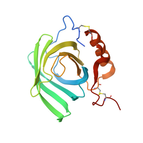
    Macromolecular crystals for X-ray diffraction studies are typically grown in vitro from pure and homogeneous samples; however, there are examples of protein crystals that have been identified in vivo. Recent developments in micro-crystallography techniques and the advent of X-ray free-electron lasers have allowed the determination of several protein structures from crystals grown in cellulo. Here, an atomic resolution (1.2 Å) crystal structure is reported of heterogeneous milk proteins grown inside a living organism in their functional niche. These in vivo-grown crystals were isolated from the midgut of an embryo within the only known viviparous cockroach, Diploptera punctata. The milk proteins crystallized in space group P1, and a structure was determined by anomalous dispersion from the native S atoms. The data revealed glycosylated proteins that adopt a lipocalin fold, bind lipids and organize to form a tightly packed crystalline lattice. A single crystal is estimated to contain more than three times the energy of an equivalent mass of dairy milk. This unique storage form of nourishment for developing embryos allows access to a constant supply of complete nutrients. Notably, the crystalline cockroach-milk proteins are highly heterogeneous with respect to amino-acid sequence, glycosylation and bound fatty-acid composition. These data present a unique example of protein heterogeneity within a single in vivo-grown crystal of a natural protein in its native environment at atomic resolution.


  • Organizational Affiliation

    Institute of Stem Cell Biology and Regenerative Medicine , Bellary Road, GKVK Campus, Bangalore, Karnataka 560 065, India.


Macromolecules
Find similar proteins by:  (by identity cutoff)  |  3D Structure
Entity ID: 1
MoleculeChains Sequence LengthOrganismDetailsImage
Milk protein164Diploptera punctataMutation(s): 0 
UniProt
Find proteins for Q6SVB6 (Diploptera punctata)
Explore Q6SVB6 
Go to UniProtKB:  Q6SVB6
Entity Groups  
Sequence Clusters30% Identity50% Identity70% Identity90% Identity95% Identity100% Identity
UniProt GroupQ6SVB6
Sequence Annotations
Expand
  • Reference Sequence
Oligosaccharides

Help

Entity ID: 2
MoleculeChains Length2D Diagram Glycosylation3D Interactions
beta-D-mannopyranose-(1-4)-2-acetamido-2-deoxy-beta-D-glucopyranose-(1-4)-2-acetamido-2-deoxy-beta-D-glucopyranose
B
3N-Glycosylation
Glycosylation Resources
GlyTouCan:  G15407YE
GlyCosmos:  G15407YE
GlyGen:  G15407YE
Entity ID: 3
MoleculeChains Length2D Diagram Glycosylation3D Interactions
2-acetamido-2-deoxy-beta-D-glucopyranose-(1-4)-2-acetamido-2-deoxy-beta-D-glucopyranose
C
2N-Glycosylation
Glycosylation Resources
GlyTouCan:  G42666HT
GlyCosmos:  G42666HT
GlyGen:  G42666HT
Experimental Data & Validation

Experimental Data

  • Method: X-RAY DIFFRACTION
  • Resolution: 1.20 Å
  • R-Value Free: 0.192 
  • R-Value Work: 0.143 
  • R-Value Observed: 0.146 
  • Space Group: P 1
Unit Cell:
Length ( Å )Angle ( ˚ )
a = 32.333α = 99.07
b = 33.28β = 100.17
c = 40.136γ = 103.7
Software Package:
Software NamePurpose
SOLVEphasing
REFMACrefinement
d*TREKdata reduction
d*TREKdata scaling

Structure Validation

View Full Validation Report


Ligand Structure Quality Assessment 


Entry History 

Deposition Data

Revision History  (Full details and data files)

  • Version 1.0: 2014-01-01
    Type: Initial release
  • Version 1.1: 2016-07-20
    Changes: Database references
  • Version 1.2: 2017-11-22
    Changes: Refinement description
  • Version 1.3: 2020-01-01
    Changes: Database references, Derived calculations
  • Version 2.0: 2020-07-29
    Type: Remediation
    Reason: Carbohydrate remediation
    Changes: Atomic model, Data collection, Derived calculations, Structure summary
  • Version 2.1: 2023-11-08
    Changes: Data collection, Database references, Refinement description, Structure summary