4NWN

Computationally Designed Two-Component Self-Assembling Tetrahedral Cage T32-28


Experimental Data Snapshot

  • Method: X-RAY DIFFRACTION
  • Resolution: 4.50 Å
  • R-Value Free: 0.343 
  • R-Value Work: 0.297 
  • R-Value Observed: 0.302 

wwPDB Validation   3D Report Full Report


This is version 1.2 of the entry. See complete history


Literature

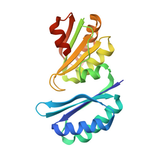
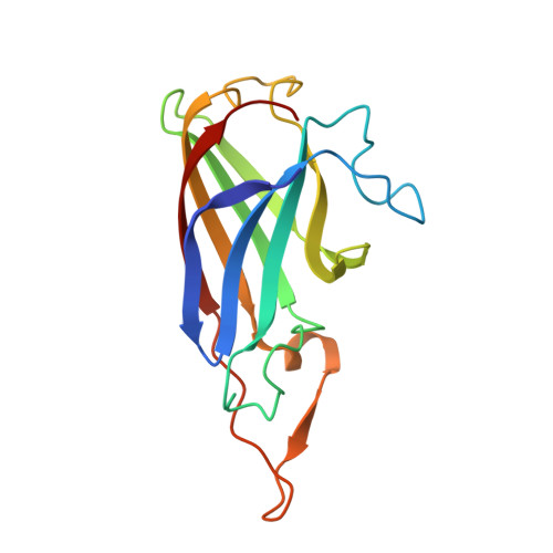
Accurate design of co-assembling multi-component protein nanomaterials.

King, N.P.Bale, J.B.Sheffler, W.McNamara, D.E.Gonen, S.Gonen, T.Yeates, T.O.Baker, D.

(2014) Nature 510: 103-108

  • DOI: https://doi.org/10.1038/nature13404
  • Primary Citation of Related Structures:  
    4NWN, 4NWO, 4NWP, 4NWQ, 4NWR

  • PubMed Abstract: 

    The self-assembly of proteins into highly ordered nanoscale architectures is a hallmark of biological systems. The sophisticated functions of these molecular machines have inspired the development of methods to engineer self-assembling protein nanostructures; however, the design of multi-component protein nanomaterials with high accuracy remains an outstanding challenge. Here we report a computational method for designing protein nanomaterials in which multiple copies of two distinct subunits co-assemble into a specific architecture. We use the method to design five 24-subunit cage-like protein nanomaterials in two distinct symmetric architectures and experimentally demonstrate that their structures are in close agreement with the computational design models. The accuracy of the method and the number and variety of two-component materials that it makes accessible suggest a route to the construction of functional protein nanomaterials tailored to specific applications.


  • Organizational Affiliation

    1] Department of Biochemistry, University of Washington, Seattle, Washington 98195, USA [2] Institute for Protein Design, University of Washington, Seattle, Washington 98195, USA [3].


Macromolecules
Find similar proteins by:  (by identity cutoff)  |  3D Structure
Entity ID: 1
MoleculeChains Sequence LengthOrganismDetailsImage
Uncharacterized protein
A, C, E, G, I
A, C, E, G, I, K, M, O, Q, S, U, W
192Campylobacter jejuni subsp. jejuni 81-176Mutation(s): 12 
Gene Names: CJE1831CJJ81176_1650PduTstm2054
Entity Groups  
Sequence Clusters30% Identity50% Identity70% Identity90% Identity95% Identity100% Identity
Sequence Annotations
Expand
  • Reference Sequence
Find similar proteins by:  (by identity cutoff)  |  3D Structure
Entity ID: 2
MoleculeChains Sequence LengthOrganismDetailsImage
Propanediol utilization: polyhedral bodies pduT
B, D, F, H, J
B, D, F, H, J, L, N, P, R, T, V, X
159Salmonella enterica subsp. enterica serovar Typhimurium str. STm5Mutation(s): 10 
Gene Names: B581_12628cjj81176_1650
UniProt
Find proteins for A0A0H3PA01 (Campylobacter jejuni subsp. jejuni serotype O:23/36 (strain 81-176))
Explore A0A0H3PA01 
Go to UniProtKB:  A0A0H3PA01
Entity Groups  
Sequence Clusters30% Identity50% Identity70% Identity90% Identity95% Identity100% Identity
UniProt GroupA0A0H3PA01
Sequence Annotations
Expand
  • Reference Sequence
Experimental Data & Validation

Experimental Data

  • Method: X-RAY DIFFRACTION
  • Resolution: 4.50 Å
  • R-Value Free: 0.343 
  • R-Value Work: 0.297 
  • R-Value Observed: 0.302 
  • Space Group: P 31 2 1
Unit Cell:
Length ( Å )Angle ( ˚ )
a = 246.01α = 90
b = 246.01β = 90
c = 290.94γ = 120
Software Package:
Software NamePurpose
XSCALEdata scaling
PHASERphasing
PHENIXrefinement
PDB_EXTRACTdata extraction
XDSdata reduction

Structure Validation

View Full Validation Report


Entry History 

Deposition Data

Revision History  (Full details and data files)

  • Version 1.0: 2014-05-28
    Type: Initial release
  • Version 1.1: 2014-06-11
    Changes: Database references
  • Version 1.2: 2023-09-20
    Changes: Data collection, Database references, Refinement description