Charge-Disproportionation Symmetry Breaking Creates a Heterodimeric Myoglobin Complex with Enhanced Affinity and Rapid Intracomplex Electron Transfer.
Trana, E.N., Nocek, J.M., Woude, J.V., Span, I., Smith, S.M., Rosenzweig, A.C., Hoffman, B.M.(2016) J Am Chem Soc 138: 12615-12628
- PubMed: 27646786 Search on PubMedSearch on PubMed Central
- DOI: https://doi.org/10.1021/jacs.6b07672
- Primary Citation Related Structures:
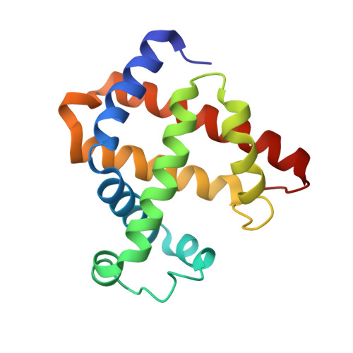
3RJ6, 4NS2, 4TWU, 4TWV - PubMed Abstract:
We report rapid photoinitiated intracomplex electron transfer (ET) within a "charge-disproportionated" myoglobin (Mb) dimer with greatly enhanced affinity. Two mutually supportive Brownian Dynamics (BD) interface redesign strategies, one a new "heme-filtering" approach, were employed to "break the symmetry" of a Mb homodimer by pairing Mb constructs with complementary highly positive and highly negative net surface charges, introduced through D/E → K and K → E mutations, respectively. BD simulations using a previously developed positive mutant, Mb(+6) = Mb(D44K/D60K/E85K), led to construction of the complementary negative mutant Mb(-6) = Mb(K45E, K63E, K95E). Simulations predict the pair will form a well-defined complex comprising a tight ensemble of conformations with nearly parallel hemes, at a metal-metal distance ∼18-19 Å. Upon expression and X-ray characterization of the partners, BD predictions were verified through ET photocycle measurements enabled by Zn-deuteroporphyrin substitution, forming the [ZnMb(-6), Fe(3+)Mb(+6)] complex. Triplet ET quenching shows charge disproportionation increases the binding constant by no less than ∼5 orders of magnitude relative to wild-type Mb values. All progress curves for charge separation (CS) and charge recombination (CR) are reproduced by a generalized kinetic model for the interprotein ET photocycle. The intracomplex ET rate constants for both CS and CR are increased by over 5 orders of magnitude, and their viscosity independence is indicative of true interprotein ET, rather than dynamic gating as seen in previous studies. The complex displays an unprecedented timecourse for CR of the CS intermediate I. After a laser flash, I forms through photoinduced CS, accumulates to a maximum concentration, then dies away through CR. However, before completely disappearing, I reappears without another flash and reaches a second maximum before disappearing completely.
- Department of Chemistry, Northwestern University , Evanston, Illinois 60208, United States.
Organizational Affiliation: