Cyanuric acid hydrolase from Azorhizobium caulinodans ORS 571: crystal structure and insights into a new class of Ser-Lys dyad proteins.
Cho, S., Shi, K., Seffernick, J.L., Dodge, A.G., Wackett, L.P., Aihara, H.(2014) PLoS One 9: e99349-e99349
- PubMed: 24915109 Search on PubMedSearch on PubMed Central
- DOI: https://doi.org/10.1371/journal.pone.0099349
- Primary Citation Related Structures:
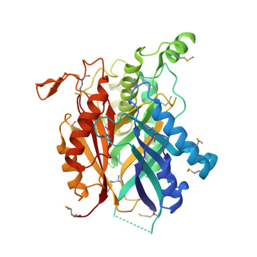
4NQ3 - PubMed Abstract:
Cyanuric acid hydrolase (CAH) catalyzes the hydrolytic ring-opening of cyanuric acid (2,4,6-trihydroxy-1,3,5-triazine), an intermediate in s-triazine bacterial degradation and a by-product from disinfection with trichloroisocyanuric acid. In the present study, an X-ray crystal structure of the CAH-barbituric acid inhibitor complex from Azorhizobium caulinodans ORS 571 has been determined at 2.7 Å resolution. The CAH protein fold consists of three structurally homologous domains forming a β-barrel-like structure with external α-helices that result in a three-fold symmetry, a dominant feature of the structure and active site that mirrors the three-fold symmetrical shape of the substrate cyanuric acid. The active site structure of CAH is similar to that of the recently determined AtzD with three pairs of active site Ser-Lys dyads. In order to determine the role of each Ser-Lys dyad in catalysis, a mutational study using a highly sensitive, enzyme-coupled assay was conducted. The 10⁹-fold loss of activity by the S226A mutant was at least ten times lower than that of the S79A and S333A mutants. In addition, bioinformatics analysis revealed the Ser226/Lys156 dyad as the only absolutely conserved dyad in the CAH/barbiturase family. These data suggest that Lys156 activates the Ser226 nucleophile which can then attack the substrate carbonyl. Our combination of structural, mutational, and bioinformatics analyses differentiates this study and provides experimental data for mechanistic insights into this unique protein family.
- Department of Biochemistry, Molecular Biology, and Biophysics, University of Minnesota, St. Paul, Minnesota, United States of America.
Organizational Affiliation: