4NK7 | pdb_00004nk7

Crystal Structure of the D. melanogaster Plk4 cryptic polo box (CPB)


Experimental Data Snapshot

  • Method: X-RAY DIFFRACTION
  • Resolution: 3.23 Å
  • R-Value Free: 
    0.264 (Depositor), 0.268 (DCC) 
  • R-Value Work: 
    0.240 (Depositor), 0.248 (DCC) 
  • R-Value Observed: 
    0.243 (Depositor) 

Starting Model: experimental
View more details

wwPDB Validation 3D Report Full Report

Validation slider image for 4NK7

This is version 1.1 of the entry. See complete history

Literature

Structure of the C. elegans ZYG-1 Cryptic Polo Box Suggests a Conserved Mechanism for Centriolar Docking of Plk4 Kinases.

Shimanovskaya, E.Viscardi, V.Lesigang, J.Lettman, M.M.Qiao, R.Svergun, D.I.Round, A.Oegema, K.Dong, G.

(2014) Structure 22: 1090-1104

  • DOI: https://doi.org/10.1016/j.str.2014.05.009
  • Primary Citation Related Structures: 
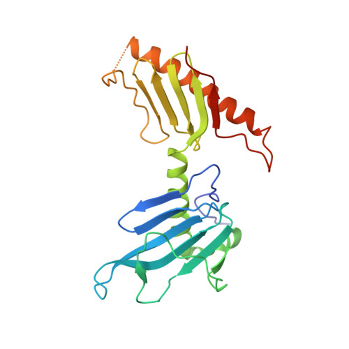
    4NK7, 4NKB

  • PubMed Abstract: 

    Plk4 family kinases control centriole assembly. Plk4s target mother centrioles through an interaction between their cryptic polo box (CPB) and acidic regions in the centriolar receptors SPD-2/Cep192 and/or Asterless/Cep152. Here, we report a crystal structure for the CPB of C. elegans ZYG-1, which forms a Z-shaped dimer containing an intermolecular β sheet with an extended basic surface patch. Biochemical and in vivo analysis revealed that electrostatic interactions dock the ZYG-1 CPB basic patch onto the SPD-2-derived acidic region to promote ZYG-1 targeting and new centriole assembly. Analysis of a different crystal form of the Drosophila Plk4 (DmPlk4) CPB suggests that it also forms a Z-shaped dimer. Comparison of the ZYG-1 and DmPlk4 CPBs revealed structural changes in the ZYG-1 CPB that confer selectivity for binding SPD-2 over Asterless-derived acidic regions. Overall, our findings suggest a conserved mechanism for centriolar docking of Plk4 homologs that initiate daughter centriole assembly.


  • Organizational Affiliation
    • Max F. Perutz Laboratories, Medical University of Vienna, 1030 Vienna, Austria.

Macromolecule Content 

  • Total Structure Weight: 25.5 kDa 
  • Atom Count: 1,705 
  • Modeled Residue Count: 212 
  • Deposited Residue Count: 223 
  • Unique protein chains: 1

Macromolecules

Find similar proteins by:|  3D Structure
Entity ID: 1
MoleculeChains  Sequence LengthOrganismDetailsImage
Serine/threonine-protein kinase PLK4223Drosophila melanogasterMutation(s): 0 
Gene Names: CG7186D. melanogaster Plk4 (residues 383-601)SAK
EC: 2.7.11.21
UniProt
Find proteins for O97143 (Drosophila melanogaster)
Explore O97143 
Go to UniProtKB:  O97143
Entity Groups
Sequence Clusters30% Identity50% Identity70% Identity90% Identity95% Identity100% Identity
UniProt GroupO97143
Sequence Annotations
Expand
Reference Sequence

Experimental Data & Validation

Experimental Data

  • Method: X-RAY DIFFRACTION
  • Resolution: 3.23 Å
  • R-Value Free:  0.264 (Depositor), 0.268 (DCC) 
  • R-Value Work:  0.240 (Depositor), 0.248 (DCC) 
  • R-Value Observed: 0.243 (Depositor) 
Space Group: P 64 2 2
Unit Cell:
Length ( Å )Angle ( ˚ )
a = 132.207α = 90
b = 132.207β = 90
c = 134.247γ = 120
Software Package:
Software NamePurpose
DNAdata collection
SOLVEphasing
PHENIXrefinement
XDSdata reduction
SCALAdata scaling

Structure Validation

View Full Validation Report


Entry History 

Deposition Data

Revision History  (Full details and data files)

  • Version 1.0: 2014-08-27
    Type: Initial release
  • Version 1.1: 2023-09-20
    Changes: Data collection, Database references, Refinement description