4NJH | pdb_00004njh

Crystal Structure of QueE from Burkholderia multivorans in complex with AdoMet and 6-carboxy-5,6,7,8-tetrahydropterin


Experimental Data Snapshot

  • Method: X-RAY DIFFRACTION
  • Resolution: 1.90 Å
  • R-Value Free: 
    0.174 (Depositor), 0.185 (DCC) 
  • R-Value Work: 
    0.147 (Depositor) 
  • R-Value Observed: 
    0.148 (Depositor) 

Starting Model: experimental
View more details

wwPDB Validation 3D Report Full Report

Validation slider image for 4NJH

Ligand Structure Quality Assessment 


This is version 1.4 of the entry. See complete history

Literature

Radical SAM enzyme QueE defines a new minimal core fold and metal-dependent mechanism.

Dowling, D.P.Bruender, N.A.Young, A.P.McCarty, R.M.Bandarian, V.Drennan, C.L.

(2014) Nat Chem Biol 10: 106-112

  • DOI: https://doi.org/10.1038/nchembio.1426
  • Primary Citation Related Structures: 
    4NJG, 4NJH, 4NJI, 4NJJ, 4NJK

  • PubMed Abstract: 

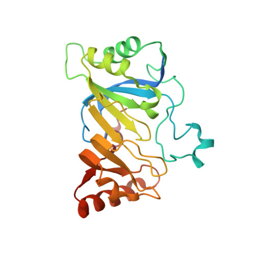
    7-carboxy-7-deazaguanine synthase (QueE) catalyzes a key S-adenosyl-L-methionine (AdoMet)- and Mg(2+)-dependent radical-mediated ring contraction step, which is common to the biosynthetic pathways of all deazapurine-containing compounds. QueE is a member of the AdoMet radical superfamily, which employs the 5'-deoxyadenosyl radical from reductive cleavage of AdoMet to initiate chemistry. To provide a mechanistic rationale for this elaborate transformation, we present the crystal structure of a QueE along with structures of pre- and post-turnover states. We find that substrate binds perpendicular to the [4Fe-4S]-bound AdoMet, exposing its C6 hydrogen atom for abstraction and generating the binding site for Mg(2+), which coordinates directly to the substrate. The Burkholderia multivorans structure reported here varies from all other previously characterized members of the AdoMet radical superfamily in that it contains a hypermodified (β6/α3) protein core and an expanded cluster-binding motif, CX14CX2C.


  • Organizational Affiliation
    • 1] Howard Hughes Medical Institute, Massachusetts Institute of Technology, Cambridge, Massachusetts, USA. [2] Department of Chemistry, Massachusetts Institute of Technology, Cambridge, Massachusetts, USA.

Macromolecule Content 

  • Total Structure Weight: 52.87 kDa 
  • Atom Count: 3,945 
  • Modeled Residue Count: 418 
  • Deposited Residue Count: 460 
  • Unique protein chains: 1

Macromolecules

Find similar proteins by:|  3D Structure
Entity ID: 1
MoleculeChains  Sequence LengthOrganismDetailsImage
7-carboxy-7-deazaguanine synthase
A, B
230Burkholderia multivorans ATCC 17616Mutation(s): 0 
Gene Names: queEBmul_3115BMULJ_00116
EC: 4.3.99.3
UniProt
Find proteins for A0A0H3KB22 (Burkholderia multivorans (strain ATCC 17616 / 249))
Explore A0A0H3KB22 
Go to UniProtKB:  A0A0H3KB22
Entity Groups
Sequence Clusters30% Identity50% Identity70% Identity90% Identity95% Identity100% Identity
UniProt GroupA0A0H3KB22
Sequence Annotations
Expand
Reference Sequence

Small Molecules

Ligands 5 Unique
IDChains Name / Formula / InChI Key2D Diagram3D Interactions
SAM

Query on SAM


Download:Ideal Coordinates CCD File
D [auth A],
I [auth B]
S-ADENOSYLMETHIONINE
C15 H22 N6 O5 S
MEFKEPWMEQBLKI-FCKMPRQPSA-N
SF4

Query on SF4


Download:Ideal Coordinates CCD File
C [auth A],
H [auth B]
IRON/SULFUR CLUSTER
Fe4 S4
LJBDFODJNLIPKO-UHFFFAOYSA-N
2K8

Query on 2K8


Download:Ideal Coordinates CCD File
E [auth A],
J [auth B]
(6R)-2-amino-4-oxo-3,4,5,6,7,8-hexahydropteridine-6-carboxylic acid
C7 H9 N5 O3
QSIYONWVWDSRRO-UWTATZPHSA-N
PO4

Query on PO4


Download:Ideal Coordinates CCD File
G [auth A],
L [auth B]
PHOSPHATE ION
O4 P
NBIIXXVUZAFLBC-UHFFFAOYSA-K
NA

Query on NA


Download:Ideal Coordinates CCD File
F [auth A],
K [auth B],
M [auth B]
SODIUM ION
Na
FKNQFGJONOIPTF-UHFFFAOYSA-N

Experimental Data & Validation

Experimental Data

  • Method: X-RAY DIFFRACTION
  • Resolution: 1.90 Å
  • R-Value Free:  0.174 (Depositor), 0.185 (DCC) 
  • R-Value Work:  0.147 (Depositor) 
  • R-Value Observed: 0.148 (Depositor) 
Space Group: P 43 21 2
Unit Cell:
Length ( Å )Angle ( ˚ )
a = 119.129α = 90
b = 119.129β = 90
c = 103.363γ = 90
Software Package:
Software NamePurpose
ADSCdata collection
PHENIXmodel building
PHENIXrefinement
HKL-2000data reduction
HKL-2000data scaling
PHENIXphasing

Structure Validation

View Full Validation Report


Ligand Structure Quality Assessment 


Entry History 

Deposition Data

Revision History  (Full details and data files)

  • Version 1.0: 2013-12-25
    Type: Initial release
  • Version 1.1: 2014-01-22
    Changes: Database references
  • Version 1.2: 2014-02-05
    Changes: Database references
  • Version 1.3: 2014-07-02
    Changes: Non-polymer description
  • Version 1.4: 2023-09-20
    Changes: Data collection, Database references, Derived calculations, Refinement description