4N7K | pdb_00004n7k

Zinc Substituted Reaction Center of the Rhodobacter sphaeroides


Experimental Data Snapshot

  • Method: X-RAY DIFFRACTION
  • Resolution: 2.85 Å
  • R-Value Free: 
    0.243 (Depositor), 0.241 (DCC) 
  • R-Value Work: 
    0.182 (Depositor), 0.182 (DCC) 
  • R-Value Observed: 
    0.185 (Depositor) 

wwPDB Validation 3D Report Full Report

Validation slider image for 4N7K

Ligand Structure Quality Assessment 


This is version 1.1 of the entry. See complete history

Literature

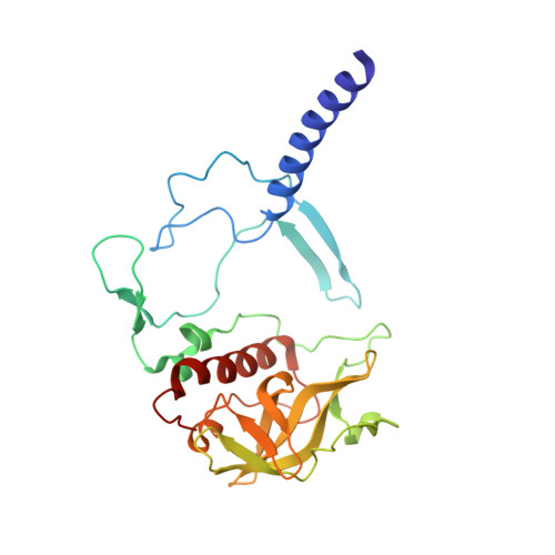
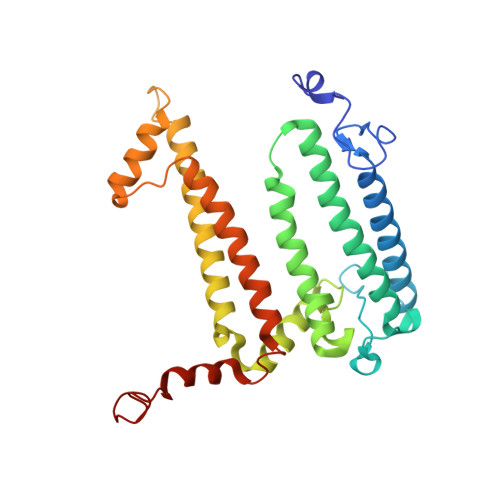
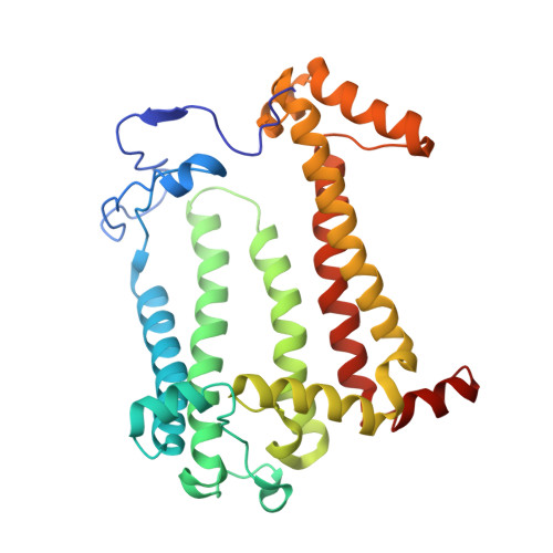
Structural and kinetic properties of Rhodobacter sphaeroides photosynthetic reaction centers containing exclusively Zn-coordinated bacteriochlorophyll as bacteriochlorin cofactors.

Saer, R.G.Pan, J.Hardjasa, A.Lin, S.Rosell, F.Mauk, A.G.Woodbury, N.W.Murphy, M.E.Beatty, J.T.

(2013) Biochim Biophys Acta 1837: 366-374

  • DOI: https://doi.org/10.1016/j.bbabio.2013.11.015
  • Primary Citation Related Structures: 
    4N7K, 4N7L

  • PubMed Abstract: 

    The Zn-BChl-containing reaction center (RC) produced in a bchD (magnesium chelatase) mutant of Rhodobacter sphaeroides assembles with six Zn-bacteriochlorophylls (Zn-BChls) in place of four Mg-containing bacteriochlorophylls (BChls) and two bacteriopheophytins (BPhes). This protein presents unique opportunities for studying biological electron transfer, as Zn-containing chlorins can exist in 4-, 5-, and (theoretically) 6-coordinate states within the RC. In this paper, the electron transfer perturbations attributed exclusively to coordination state effects are separated from those attributed to the presence, absence, or type of metal in the bacteriochlorin at the HA pocket of the RC. The presence of a 4-coordinate Zn(2+) ion in the HA bacteriochlorin instead of BPhe results in a small decrease in the rates of the P*→P(+)HA(-)→P(+)QA(-) electron transfer, and the charge separation yield is not greatly perturbed; however coordination of the Zn(2+) by a fifth ligand provided by a histidine residue results in a larger rate decrease and yield loss. We also report the first crystal structure of a Zn-BChl-containing RC, confirming that the HA Zn-BChl was either 4- or 5-coordinate in the two types of Zn-BChl-containing RCs studied here. Interestingly, a large degree of disorder, in combination with a relatively weak anomalous difference electron density was found in the HB pocket. These data, in combination with spectroscopic results, indicate partial occupancy of this binding pocket. These findings provide insights into the use of BPhe as the bacteriochlorin pigment of choice at HA in both BChl- and Zn-BChl-containing RCs found in nature.


  • Organizational Affiliation
    • Department of Microbiology and Immunology, The University of British Columbia, 2350 Health Sciences Mall, Vancouver, BC V6T 1Z3, Canada.

Macromolecule Content 

  • Total Structure Weight: 104.91 kDa 
  • Atom Count: 7,634 
  • Modeled Residue Count: 825 
  • Deposited Residue Count: 825 
  • Unique protein chains: 3

Macromolecules

Find similar proteins by:|  3D Structure
Entity ID: 1
MoleculeChains  Sequence LengthOrganismDetailsImage
Reaction Center H ChainA [auth H]241Cereibacter sphaeroidesMutation(s): 0 
Membrane Entity: Yes 
UniProt
Find proteins for P0C0Y7 (Cereibacter sphaeroides)
Explore P0C0Y7 
Go to UniProtKB:  P0C0Y7
Entity Groups
Sequence Clusters30% Identity50% Identity70% Identity90% Identity95% Identity100% Identity
UniProt GroupP0C0Y7
Sequence Annotations
Expand
Reference Sequence
Find similar proteins by:|  3D Structure
Entity ID: 2
MoleculeChains  Sequence LengthOrganismDetailsImage
Reaction center protein L chainB [auth L]281Cereibacter sphaeroidesMutation(s): 0 
Gene Names: pufL
Membrane Entity: Yes 
UniProt
Find proteins for P0C0Y8 (Cereibacter sphaeroides)
Explore P0C0Y8 
Go to UniProtKB:  P0C0Y8
Entity Groups
Sequence Clusters30% Identity50% Identity70% Identity90% Identity95% Identity100% Identity
UniProt GroupP0C0Y8
Sequence Annotations
Expand
Reference Sequence
Find similar proteins by:|  3D Structure
Entity ID: 3
MoleculeChains  Sequence LengthOrganismDetailsImage
Reaction center protein M chainC [auth M]303Cereibacter sphaeroidesMutation(s): 0 
Gene Names: pufM
Membrane Entity: Yes 
UniProt
Find proteins for P0C0Y9 (Cereibacter sphaeroides)
Explore P0C0Y9 
Go to UniProtKB:  P0C0Y9
Entity Groups
Sequence Clusters30% Identity50% Identity70% Identity90% Identity95% Identity100% Identity
UniProt GroupP0C0Y9
Sequence Annotations
Expand
Reference Sequence

Small Molecules

Ligands 11 Unique
IDChains Name / Formula / InChI Key2D Diagram3D Interactions
CDL

Query on CDL


Download:Ideal Coordinates CCD File
DA [auth M]CARDIOLIPIN
C81 H156 O17 P2
XVTUQDWPJJBEHJ-KZCWQMDCSA-L
GGD

Query on GGD


Download:Ideal Coordinates CCD File
H
NONADEC-10-ENOIC ACID 2-[3,4-DIHYDROXY-6-HYDROXYMETHYL-5-(3,4,5-TRIHYDROXY-6-HYDROXYMETHYL-TETRAHYDRO-PYRAN-2-YLOXY)-TETRAHYDRO-PYRAN-2-YLOXY] -1-OCTADEC-9-ENOYLOXYMETHYL-ETHYL ESTER
C52 H94 O15
OGKQOSUHVSQNSH-IQXSWRQFSA-N
2GO

Query on 2GO


Download:Ideal Coordinates CCD File
AA [auth M]
I [auth L]
M [auth L]
O [auth L]
V [auth M]
AA [auth M],
I [auth L],
M [auth L],
O [auth L],
V [auth M],
W [auth M]
[methyl 9-acetyl-14-ethyl-20-hydroxy-4,8,13,18-tetramethyl-3-{3-oxo-3-[(3,7,11,15-tetramethylhexadec-2-en-1-yl)oxy]propyl}-3,4,20,21-tetradehydrophorbine-21-carboxylatato(2-)-kappa~4~N~23~,N~24~,N~25~,N~26~]zinc
C55 H70 N4 O6 Zn
WIUVDOQKCIQUQM-HCIDKWDRSA-L
U10

Query on U10


Download:Ideal Coordinates CCD File
BA [auth M],
N [auth L]
UBIQUINONE-10
C59 H90 O4
ACTIUHUUMQJHFO-UPTCCGCDSA-N
PC1

Query on PC1


Download:Ideal Coordinates CCD File
EA [auth M]1,2-DIACYL-SN-GLYCERO-3-PHOSPHOCHOLINE
C44 H88 N O8 P
NRJAVPSFFCBXDT-HUESYALOSA-N
SPO

Query on SPO


Download:Ideal Coordinates CCD File
CA [auth M]SPHEROIDENE
C41 H60 O
FJOCMTHZSURUFA-KXCOHNEYSA-N
LDA

Query on LDA


Download:Ideal Coordinates CCD File
J [auth L],
K [auth L],
L,
X [auth M],
Y [auth M]
LAURYL DIMETHYLAMINE-N-OXIDE
C14 H31 N O
SYELZBGXAIXKHU-UHFFFAOYSA-N
HTO

Query on HTO


Download:Ideal Coordinates CCD File
Q [auth L],
R [auth L]
HEPTANE-1,2,3-TRIOL
C7 H16 O3
HXYCHJFUBNTKQR-RNFRBKRXSA-N
PO4

Query on PO4


Download:Ideal Coordinates CCD File
P [auth L]PHOSPHATE ION
O4 P
NBIIXXVUZAFLBC-UHFFFAOYSA-K
GOL

Query on GOL


Download:Ideal Coordinates CCD File
D [auth H]
E [auth H]
F [auth H]
G [auth H]
S [auth L]
D [auth H],
E [auth H],
F [auth H],
G [auth H],
S [auth L],
T [auth L],
U [auth L]
GLYCEROL
C3 H8 O3
PEDCQBHIVMGVHV-UHFFFAOYSA-N
FE

Query on FE


Download:Ideal Coordinates CCD File
Z [auth M]FE (III) ION
Fe
VTLYFUHAOXGGBS-UHFFFAOYSA-N

Experimental Data & Validation

Experimental Data

  • Method: X-RAY DIFFRACTION
  • Resolution: 2.85 Å
  • R-Value Free:  0.243 (Depositor), 0.241 (DCC) 
  • R-Value Work:  0.182 (Depositor), 0.182 (DCC) 
  • R-Value Observed: 0.185 (Depositor) 
Space Group: P 31 2 1
Unit Cell:
Length ( Å )Angle ( ˚ )
a = 139.45α = 90
b = 139.45β = 90
c = 184.181γ = 120
Software Package:
Software NamePurpose
SCALAdata scaling
REFMACrefinement
PDB_EXTRACTdata extraction

Structure Validation

View Full Validation Report


Ligand Structure Quality Assessment 


Entry History 

Deposition Data

Revision History  (Full details and data files)

  • Version 1.0: 2014-02-05
    Type: Initial release
  • Version 1.1: 2024-02-28
    Changes: Data collection, Database references, Derived calculations, Structure summary