4N7B

Structure of the E-1-hydroxy-2-methyl-but-2-enyl-4-diphosphate reductase from Plasmodium falciparum


Experimental Data Snapshot

  • Method: X-RAY DIFFRACTION
  • Resolution: 2.20 Å
  • R-Value Free: 0.195 
  • R-Value Work: 0.172 
  • R-Value Observed: 0.173 

wwPDB Validation   3D Report Full Report


This is version 1.3 of the entry. See complete history


Literature

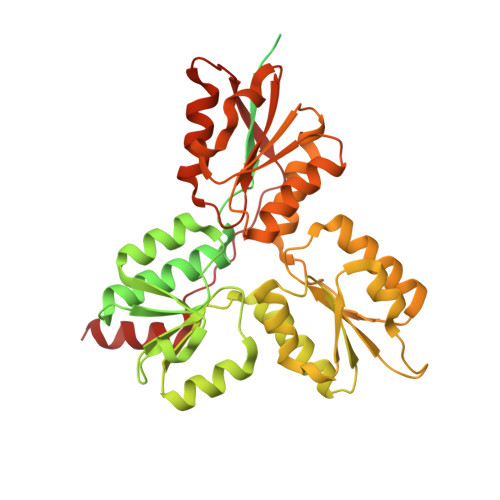
Structure of the (E)-4-hydroxy-3-methyl-but-2-enyl-diphosphate reductase from Plasmodium falciparum.

Rekittke, I.Olkhova, E.Wiesner, J.Demmer, U.Warkentin, E.Jomaa, H.Ermler, U.

(2013) FEBS Lett 587: 3968-3972

  • DOI: https://doi.org/10.1016/j.febslet.2013.10.029
  • Primary Citation of Related Structures:  
    4N7B

  • PubMed Abstract: 

    Terpenoid precursor biosynthesis occurs in human and many pathogenic organisms via the mevalonate and 2-C-methyl-d-erythritol-4-phosphate (MEP) pathways, respectively. We determined the X-ray structure of the Fe/S containing (E)-4-hydroxy-3-methyl-but-2-enyl-diphosphate reductase (LytB) of the pathogenic protozoa Plasmodium falciparum which catalyzes the terminal step of the MEP pathway. The cloverleaf fold and the active site of P. falciparum LytB corresponds to those of the Aquifex aeolicus and Escherichia coli enzymes. Its distinct electron donor [2Fe-2S] ferredoxin was modeled to its binding site by docking calculations. The presented structural data provide a platform for a rational search of anti-malarian drugs.


  • Organizational Affiliation

    Medizinische Klinik IV (Hämatologie), Justus-Liebig-Universität Giessen, Klinikstrasse 33, D-35392 Giessen, Germany.


Macromolecules
Find similar proteins by:  (by identity cutoff)  |  3D Structure
Entity ID: 1
MoleculeChains Sequence LengthOrganismDetailsImage
LytB541Plasmodium falciparumMutation(s): 0 
Gene Names: lytB
EC: 1.17.7.4
UniProt
Find proteins for Q8I295 (Plasmodium falciparum (isolate 3D7))
Explore Q8I295 
Go to UniProtKB:  Q8I295
Entity Groups  
Sequence Clusters30% Identity50% Identity70% Identity90% Identity95% Identity100% Identity
UniProt GroupQ8I295
Sequence Annotations
Expand
  • Reference Sequence
Small Molecules
Ligands 4 Unique
IDChains Name / Formula / InChI Key2D Diagram3D Interactions
F3S
Query on F3S

Download Ideal Coordinates CCD File 
B [auth A]FE3-S4 CLUSTER
Fe3 S4
FCXHZBQOKRZXKS-UHFFFAOYSA-N
SO4
Query on SO4

Download Ideal Coordinates CCD File 
C [auth A],
D [auth A]
SULFATE ION
O4 S
QAOWNCQODCNURD-UHFFFAOYSA-L
GOL
Query on GOL

Download Ideal Coordinates CCD File 
E [auth A]GLYCEROL
C3 H8 O3
PEDCQBHIVMGVHV-UHFFFAOYSA-N
NA
Query on NA

Download Ideal Coordinates CCD File 
F [auth A],
G [auth A]
SODIUM ION
Na
FKNQFGJONOIPTF-UHFFFAOYSA-N
Experimental Data & Validation

Experimental Data

  • Method: X-RAY DIFFRACTION
  • Resolution: 2.20 Å
  • R-Value Free: 0.195 
  • R-Value Work: 0.172 
  • R-Value Observed: 0.173 
  • Space Group: P 31 2 1
Unit Cell:
Length ( Å )Angle ( ˚ )
a = 115.96α = 90
b = 115.96β = 90
c = 76.93γ = 120
Software Package:
Software NamePurpose
SHARPphasing
PHENIXrefinement
XDSdata reduction
XSCALEdata scaling

Structure Validation

View Full Validation Report


Entry History 

Deposition Data

Revision History  (Full details and data files)

  • Version 1.0: 2013-11-20
    Type: Initial release
  • Version 1.1: 2013-12-18
    Changes: Database references
  • Version 1.2: 2017-11-15
    Changes: Refinement description
  • Version 1.3: 2024-02-28
    Changes: Data collection, Database references, Derived calculations