First Structural Insights into alpha-L-Arabinofuranosidases from the Two GH62 Glycoside Hydrolase Subfamilies.
Siguier, B., Haon, M., Nahoum, V., Marcellin, M., Burlet-Schiltz, O., Coutinho, P.M., Henrissat, B., Mourey, L., O'Donohue, M.J., Berrin, J.G., Tranier, S., Dumon, C.(2014) J Biol Chem 289: 5261-5273
- PubMed: 24394409 Search on PubMedSearch on PubMed Central
- DOI: https://doi.org/10.1074/jbc.M113.528133
- Primary Citation Related Structures:
4N1I, 4N2R, 4N2Z, 4N4B - PubMed Abstract:
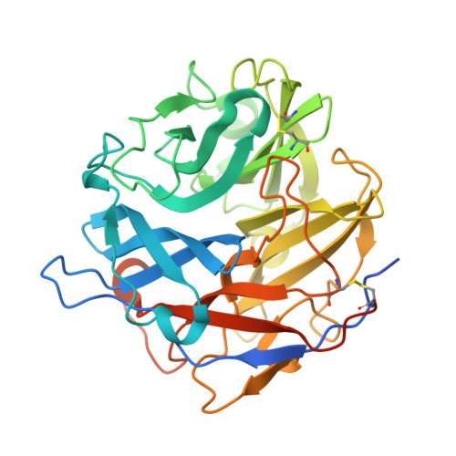
α-L-arabinofuranosidases are glycoside hydrolases that specifically hydrolyze non-reducing residues from arabinose-containing polysaccharides. In the case of arabinoxylans, which are the main components of hemicellulose, they are part of microbial xylanolytic systems and are necessary for complete breakdown of arabinoxylans. Glycoside hydrolase family 62 (GH62) is currently a small family of α-L-arabinofuranosidases that contains only bacterial and fungal members. Little is known about the GH62 mechanism of action, because only a few members have been biochemically characterized and no three-dimensional structure is available. Here, we present the first crystal structures of two fungal GH62 α-L-arabinofuranosidases from the basidiomycete Ustilago maydis (UmAbf62A) and ascomycete Podospora anserina (PaAbf62A). Both enzymes are able to efficiently remove the α-L-arabinosyl substituents from arabinoxylan. The overall three-dimensional structure of UmAbf62A and PaAbf62A reveals a five-bladed β-propeller fold that confirms their predicted classification into clan GH-F together with GH43 α-L-arabinofuranosidases. Crystallographic structures of the complexes with arabinose and cellotriose reveal the important role of subsites +1 and +2 for sugar binding. Intriguingly, we observed that PaAbf62A was inhibited by cello-oligosaccharides and displayed binding affinity to cellulose although no activity was observed on a range of cellulosic substrates. Bioinformatic analyses showed that UmAbf62A and PaAbf62A belong to two distinct subfamilies within the GH62 family. The results presented here provide a framework to better investigate the structure-function relationships within the GH62 family.
- From the Université de Toulouse, INSA, UPS, INP, LISBP, 135 Avenue de Rangueil, F-31077 Toulouse.
Organizational Affiliation: