4MXJ

Thermolysin in complex with UBTLN35


Experimental Data Snapshot

  • Method: X-RAY DIFFRACTION
  • Resolution: 1.35 Å
  • R-Value Free: 0.151 
  • R-Value Work: 0.116 
  • R-Value Observed: 0.118 

wwPDB Validation   3D Report Full Report


Ligand Structure Quality Assessment 


This is version 1.2 of the entry. See complete history


Literature

Methyl, Ethyl, Propyl, Butyl: Futile But Not for Water, as the Correlation of Structure and Thermodynamic Signature Shows in a Congeneric Series of Thermolysin Inhibitors.

Krimmer, S.G.Betz, M.Heine, A.Klebe, G.

(2014) ChemMedChem 4: 833-846

  • DOI: https://doi.org/10.1002/cmdc.201400013
  • Primary Citation of Related Structures:  
    4MTW, 4MWP, 4MXJ, 4MZN, 4N4E, 4N5P, 4N66, 4OI5

  • PubMed Abstract: 

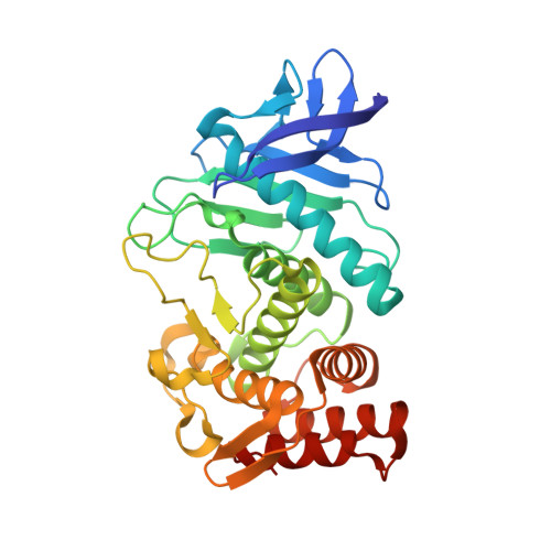
    Water is ubiquitously present in any biological system and has therefore to be regarded as an additional binding partner in the protein-ligand binding process. Upon complex formation, a new solvent-exposed surface is generated and water molecules from the first solvation layer will arrange around this newly formed surface. So far, the influence of such water arrangements on the ligand binding properties is unknown. In this study, the binding modes of nine congeneric phosphonamidate-type inhibitors with systematically varied, size-increasing hydrophobic P2 ' substituents (from methyl to phenylethyl) addressing the hydrophobic, solvent-exposed S2 ' pocket of thermolysin were analyzed by high-resolution crystal structures and correlated with their thermodynamic binding profiles as measured by isothermal titration calorimetry. Overall, ΔΔG spreads over 7.0 kJ mol(-1) , ΔΔH varies by 15.8 kJ mol(-1) , and -TΔΔS by 12.1 kJ mol(-1) . Throughout the series, these changes correlate remarkably well with the geometric differences of water molecules arranged adjacent to the P2 ' substituents. Ligands with medium-sized P2 ' substituents exhibit highest affinities, presumably because of their optimal solvation patterns around these complexes. The addition, removal, or rearrangement of even a single methyl group can result in a strong modulation of the adjacent water network pattern shifting from enthalpy to entropy-driven binding. In conclusion, the quality of a water network assembled around a protein-ligand complex influences the enthalpy/entropy signature and can even modulate affinity to a surprising extent.


  • Organizational Affiliation

    Department of Pharmaceutical Chemistry, University of Marburg, Marbacher Weg 6, 35032 Marburg (Germany).


Macromolecules
Find similar proteins by:  (by identity cutoff)  |  3D Structure
Entity ID: 1
MoleculeChains Sequence LengthOrganismDetailsImage
ThermolysinA [auth E]316Bacillus thermoproteolyticusMutation(s): 0 
EC: 3.4.24.27
UniProt
Find proteins for P00800 (Bacillus thermoproteolyticus)
Explore P00800 
Go to UniProtKB:  P00800
Entity Groups  
Sequence Clusters30% Identity50% Identity70% Identity90% Identity95% Identity100% Identity
UniProt GroupP00800
Sequence Annotations
Expand
  • Reference Sequence
Small Molecules
Ligands 5 Unique
IDChains Name / Formula / InChI Key2D Diagram3D Interactions
2G8
Query on 2G8

Download Ideal Coordinates CCD File 
L [auth E]P-((((benzyloxy)carbonyl)amino)methyl)-N-((S)-4-methyl-1-oxo-1-(propylamino)pentan-2-yl)phosphonamidic acid
C18 H30 N3 O5 P
GSTDCDKICCYPKA-INIZCTEOSA-N
GOL
Query on GOL

Download Ideal Coordinates CCD File 
G [auth E],
K [auth E]
GLYCEROL
C3 H8 O3
PEDCQBHIVMGVHV-UHFFFAOYSA-N
DMS
Query on DMS

Download Ideal Coordinates CCD File 
H [auth E],
I [auth E],
J [auth E]
DIMETHYL SULFOXIDE
C2 H6 O S
IAZDPXIOMUYVGZ-UHFFFAOYSA-N
ZN
Query on ZN

Download Ideal Coordinates CCD File 
B [auth E]ZINC ION
Zn
PTFCDOFLOPIGGS-UHFFFAOYSA-N
CA
Query on CA

Download Ideal Coordinates CCD File 
C [auth E],
D [auth E],
E,
F [auth E]
CALCIUM ION
Ca
BHPQYMZQTOCNFJ-UHFFFAOYSA-N
Experimental Data & Validation

Experimental Data

  • Method: X-RAY DIFFRACTION
  • Resolution: 1.35 Å
  • R-Value Free: 0.151 
  • R-Value Work: 0.116 
  • R-Value Observed: 0.118 
  • Space Group: P 61 2 2
Unit Cell:
Length ( Å )Angle ( ˚ )
a = 92.567α = 90
b = 92.567β = 90
c = 130.779γ = 120
Software Package:
Software NamePurpose
MAR345dtbdata collection
PHASERphasing
PHENIXrefinement
HKL-2000data reduction
HKL-2000data scaling

Structure Validation

View Full Validation Report


Ligand Structure Quality Assessment 


Entry History 

Deposition Data

Revision History  (Full details and data files)

  • Version 1.0: 2014-04-02
    Type: Initial release
  • Version 1.1: 2014-04-16
    Changes: Database references
  • Version 1.2: 2023-09-20
    Changes: Data collection, Database references, Derived calculations, Refinement description