4MXI | pdb_00004mxi

ClpP Ser98dhA


Experimental Data Snapshot

  • Method: X-RAY DIFFRACTION
  • Resolution: 2.30 Å
  • R-Value Free: 
    0.221 (Depositor), 0.219 (DCC) 
  • R-Value Work: 
    0.196 (Depositor), 0.198 (DCC) 
  • R-Value Observed: 
    0.197 (Depositor) 

Starting Model: experimental
View more details

wwPDB Validation 3D Report Full Report

Validation slider image for 4MXI

This is version 1.2 of the entry. See complete history

Literature

Disruption of Oligomerization and Dehydroalanine Formation as Mechanisms for ClpP Protease Inhibition.

Gersch, M.Kolb, R.Alte, F.Groll, M.Sieber, S.A.

(2014) J Am Chem Soc 136: 1360-1366

  • DOI: https://doi.org/10.1021/ja4082793
  • Primary Citation Related Structures: 
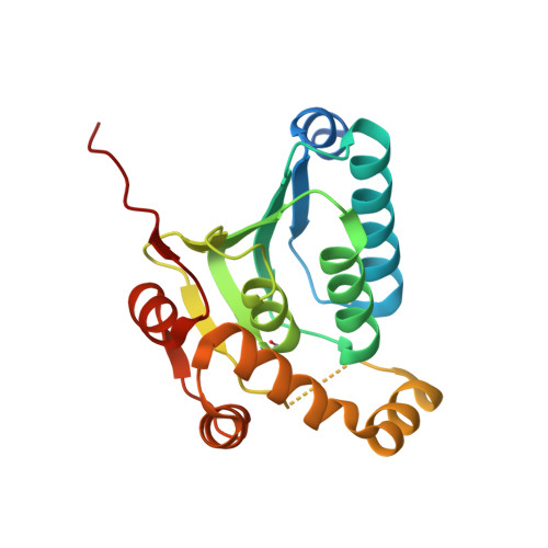
    4MXI

  • PubMed Abstract: 

    Over 100 protease inhibitors are currently used in the clinics, and most of them use blockage of the active site for their mode of inhibition. Among the protease drug targets are several enzymes for which the correct multimeric assembly is crucial to their activity, such as the proteasome and the HIV protease. Here, we present a novel mechanism of protease inhibition that relies on active-site-directed small molecules that disassemble the protease complex. We show the applicability of this mechanism within the ClpP protease family, whose members are tetradecameric serine proteases and serve as regulators of several cellular processes, including homeostasis and virulence. Compound binding to ClpP in a substoichiometric fashion triggers the formation of completely inactive heptamers. Moreover, we report the selective β-sultam-induced dehydroalanine formation of the active site serine. This reaction proceeds through sulfonylation and subsequent elimination, thereby obliterating the catalytic charge relay system. The identity of the dehydroalanine was confirmed by mass spectrometry and crystallography. Activity-based protein profiling experiments suggest the formation of a dehydroalanine moiety in living S. aureus cells upon β-sultam treatment. Collectively, these findings extend our view on multicomponent protease inhibition that until now has mainly relied on blockage of the active site or occupation of a regulatory allosteric site.


  • Organizational Affiliation
    • Center for Integrated Protein Science at the Department of Chemistry, Institute of Advanced Studies IAS and ‡Center for Integrated Protein Science at the Department of Chemistry, Technische Universität München , Lichtenbergstrasse 4, Garching D-85747, Germany.

Macromolecule Content 

  • Total Structure Weight: 150.63 kDa 
  • Atom Count: 9,634 
  • Modeled Residue Count: 1,184 
  • Deposited Residue Count: 1,365 
  • Unique protein chains: 1

Macromolecules

Find similar proteins by:|  3D Structure
Entity ID: 1
MoleculeChains  Sequence LengthOrganismDetailsImage
ATP-dependent Clp protease proteolytic subunit
A, B, C, D, E
A, B, C, D, E, F, G
195Staphylococcus aureus subsp. aureus NCTC 8325Mutation(s): 0 
Gene Names: clpPSAOUHSC_00790
EC: 3.4.21.92
UniProt
Find proteins for Q2G036 (Staphylococcus aureus (strain NCTC 8325 / PS 47))
Explore Q2G036 
Go to UniProtKB:  Q2G036
Entity Groups
Sequence Clusters30% Identity50% Identity70% Identity90% Identity95% Identity100% Identity
UniProt GroupQ2G036
Sequence Annotations
Expand
Reference Sequence

Small Molecules

Modified Residues  1 Unique
IDChains TypeFormula2D DiagramParent
DHA
Query on DHA
A, B, C, D, E
A, B, C, D, E, F, G
PEPTIDE LINKINGC3 H5 N O2SER

Experimental Data & Validation

Experimental Data

  • Method: X-RAY DIFFRACTION
  • Resolution: 2.30 Å
  • R-Value Free:  0.221 (Depositor), 0.219 (DCC) 
  • R-Value Work:  0.196 (Depositor), 0.198 (DCC) 
  • R-Value Observed: 0.197 (Depositor) 
Space Group: P 61 2 2
Unit Cell:
Length ( Å )Angle ( ˚ )
a = 121.59α = 90
b = 121.59β = 90
c = 402.95γ = 120
Software Package:
Software NamePurpose
XDSdata scaling
PHASERphasing
REFMACrefinement
XDSdata reduction
XSCALEdata scaling

Structure Validation

View Full Validation Report


Entry History 

Deposition Data

Revision History  (Full details and data files)

  • Version 1.0: 2013-10-23
    Type: Initial release
  • Version 1.1: 2014-02-12
    Changes: Database references
  • Version 1.2: 2023-09-20
    Changes: Data collection, Database references, Derived calculations, Refinement description