


X-ray structure of trypsin-inhibitor-complex
Wagner, S., Heine, A., Steinmetzer, T.To be published.
Experimental Data Snapshot
Starting Model: experimental
View more details
Entity ID: 1 | |||||
|---|---|---|---|---|---|
| Molecule | Chains | Sequence Length | Organism | Details | Image |
| Cationic trypsin | 223 | Bos taurus | Mutation(s): 0 EC: 3.4.21.4 | 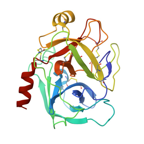 | |
UniProt | |||||
Entity Groups | |||||
| Sequence Clusters | 30% Identity50% Identity70% Identity90% Identity95% Identity100% Identity | ||||
| UniProt Group | P00760 | ||||
Sequence AnnotationsExpand | |||||
Reference Sequence | |||||
| Ligands 3 Unique | |||||
|---|---|---|---|---|---|
| ID | Chains | Name / Formula / InChI Key | 2D Diagram | 3D Interactions | |
| BZY Download:Ideal Coordinates CCD File | C [auth A] | (2R)-2-amino-N-{(2S)-1-[(4-carbamimidoylbenzyl)amino]-1-oxopropan-2-yl}-4-(4-hydroxyphenyl)butanamide C21 H27 N5 O3 GVEMEQSTGJKMAX-SCLBCKFNSA-N |  | ||
| IMD Download:Ideal Coordinates CCD File | D [auth A] | IMIDAZOLE C3 H5 N2 RAXXELZNTBOGNW-UHFFFAOYSA-O |  | ||
| CA Download:Ideal Coordinates CCD File | B [auth A] | CALCIUM ION Ca BHPQYMZQTOCNFJ-UHFFFAOYSA-N |  | ||
| Length ( Å ) | Angle ( ˚ ) |
|---|---|
| a = 54.959 | α = 90 |
| b = 54.959 | β = 90 |
| c = 108.059 | γ = 120 |
| Software Name | Purpose |
|---|---|
| MAR345dtb | data collection |
| PHASER | phasing |
| PHENIX | refinement |
| HKL-2000 | data reduction |
| HKL-2000 | data scaling |


