Comparative study of two GH19 chitinase-like proteins from Hevea brasiliensis, one exhibiting a novel carbohydrate-binding domain.
Martinez-Caballero, S., Cano-Sanchez, P., Mares-Mejia, I., Diaz-Sanchez, A.G., Macias-Rubalcava, M.L., Hermoso, J.A., Rodriguez-Romero, A.(2014) FEBS J 281: 4535-4554
- PubMed: 25104038 Search on PubMed
- DOI: https://doi.org/10.1111/febs.12962
- Primary Citation Related Structures:
4MPI, 4MST - PubMed Abstract:
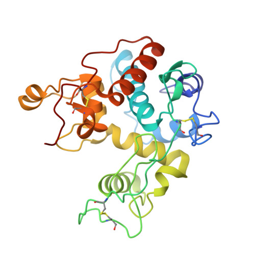
Plants express chitinase and chitinase-like proteins (CLPs) belonging to the glycosyl hydrolases of the GH18 and GH19 families, which exhibit varied functions. CLPs in the GH18 family have been structurally and functionally characterized; however, there are no structures available for any member of the GH19 family. In this study, two CLPs of the GH19 family from the rubber tree Hevea brasiliensis (HbCLP1 and HbCLP2) were cloned, expressed and characterized. HbCLP1 was identical to the allergen Hev b 11.0101 previously described by others, while HbCLP2 was a novel isoform exhibiting an unusual half chitin-binding domain before the catalytic domain. Sequence alignments showed that in the two proteins the catalytic residues Glu117 and Glu147 in HbCLP1 and HbCLP2, respectively, were mutated to Ala, accounting for the lack of activity. Nonetheless, both CLPs bound chitin and chitotriose (GlcNAc)3 with high affinities, as evaluated with chitin-affinity chromatography and tryptophan fluorescence experiments. The chitin-binding domains also bound chitotriose with even higher affinities. The crystal structures of the HbCLP1-isolated domains were determined at high resolution. The analysis of the crystallographic models and docking experiments using (GlcNAc)6 oligosaccharides provides evidence of the residues involved in sugar binding. Endochitinase activity was restored in both proteins by mutating residues A117E (HbCLP1) and A147E (HbCLP2); the distance between the catalytic proton donor and the catalytic nucleophile in the in silico mutated residues was 9.5 Å, as occurs in inverting enzymes. HbCLP1 and HbCLP2 were highly thermostable and exhibited antifungal activity against Alternaria alternata, suggesting their participation in plant defense mechanisms.
- Instituto de Química, Universidad Nacional Autónoma de México, Ciudad Universitaria, México.
Organizational Affiliation: