4MMN | pdb_00004mmn

Structural and biochemical analysis of type II free methionine-R-sulfoxide reductase from Thermoplasma acidophilum


Experimental Data Snapshot

  • Method: X-RAY DIFFRACTION
  • Resolution: 2.60 Å
  • R-Value Free: 
    0.240 (Depositor), 0.241 (DCC) 
  • R-Value Work: 
    0.196 (Depositor), 0.198 (DCC) 
  • R-Value Observed: 
    0.197 (Depositor) 

Starting Model: experimental
View more details

wwPDB Validation 3D Report Full Report

Validation slider image for 4MMN

This is version 1.2 of the entry. See complete history

Literature

Structural and biochemical analysis of a type II free methionine-R-sulfoxide reductase from Thermoplasma acidophilum

Kim, H.S.Kwak, G.H.Lee, K.Jo, C.H.Hwang, K.Y.Kim, H.Y.

(2014) Arch Biochem Biophys 560: 10-19

  • DOI: https://doi.org/10.1016/j.abb.2014.07.009
  • Primary Citation Related Structures: 
    4MMN, 4MN7

  • PubMed Abstract: 

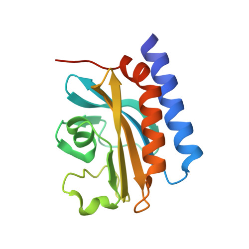
    Free methionine-R-sulfoxide reductase (fRMsr) enzymes only reduce the free form of methionine-R-sulfoxide and can be grouped into two types with respect to the number of conserved Cys residues in the active sites. In this work, the crystal structures of type II fRMsr from Thermoplasma acidophilum (TafRMsr), which contains two conserved Cys, have been determined in native form and in a complex with the substrate. The overall structure of TafRMsr consists of a central β-sheet encompassed by a two-α-helix bundle flanking on one side and one small α-helix on the other side. Based on biochemical and growth complementation assays, Cys(84) is demonstrated to be the catalytic Cys. The data also show that TafRMsr functions as an antioxidant protein. Structural analyses reveal insights into substrate recognition and orientation, conformational changes in the active site during substrate binding, and the role of active site residues in substrate binding. A model for the catalytic mechanism of type II TafRMsr is suggested, in which intramolecular disulfide bond formation is not involved. In addition, the biochemical, enzymatic, and structural properties of type II TafRMsr are compared with those of type I enzymes.


  • Organizational Affiliation
    • Department of Biosystems and Biotechnology, College of Life Sciences and Biotechnology, Korea University, Seoul 136-701, Republic of Korea.

Macromolecule Content 

  • Total Structure Weight: 136.11 kDa 
  • Atom Count: 9,089 
  • Modeled Residue Count: 1,124 
  • Deposited Residue Count: 1,192 
  • Unique protein chains: 1

Macromolecules

Find similar proteins by:|  3D Structure
Entity ID: 1
MoleculeChains  Sequence LengthOrganismDetailsImage
Putative uncharacterized protein Ta0848
A, B, C, D, E
A, B, C, D, E, F, G, H
149Thermoplasma acidophilum DSM 1728Mutation(s): 0 
Gene Names: Ta0848
UniProt
Find proteins for Q9HJW5 (Thermoplasma acidophilum (strain ATCC 25905 / DSM 1728 / JCM 9062 / NBRC 15155 / AMRC-C165))
Explore Q9HJW5 
Go to UniProtKB:  Q9HJW5
Entity Groups
Sequence Clusters30% Identity50% Identity70% Identity90% Identity95% Identity100% Identity
UniProt GroupQ9HJW5
Sequence Annotations
Expand
Reference Sequence

Experimental Data & Validation

Experimental Data

  • Method: X-RAY DIFFRACTION
  • Resolution: 2.60 Å
  • R-Value Free:  0.240 (Depositor), 0.241 (DCC) 
  • R-Value Work:  0.196 (Depositor), 0.198 (DCC) 
  • R-Value Observed: 0.197 (Depositor) 
Space Group: H 3
Unit Cell:
Length ( Å )Angle ( ˚ )
a = 158.902α = 90
b = 158.902β = 90
c = 194.596γ = 120
Software Package:
Software NamePurpose
HKL-2000data collection
PHENIXrefinement
HKL-2000data reduction
HKL-2000data scaling

Structure Validation

View Full Validation Report


Entry History 

Deposition Data

Revision History  (Full details and data files)

  • Version 1.0: 2014-08-06
    Type: Initial release
  • Version 1.1: 2014-12-17
    Changes: Database references
  • Version 1.2: 2023-11-08
    Changes: Data collection, Database references, Refinement description