4MIH

Pyranose 2-oxidase from Phanerochaete chrysosporium, recombinant H158A mutant


Experimental Data Snapshot

  • Method: X-RAY DIFFRACTION
  • Resolution: 2.40 Å
  • R-Value Free: 0.265 
  • R-Value Work: 0.205 
  • R-Value Observed: 0.206 

wwPDB Validation   3D Report Full Report


Ligand Structure Quality Assessment 


This is version 1.3 of the entry. See complete history


Literature

Crystal structures of Phanerochaete chrysosporium pyranose 2-oxidase suggest that the N-terminus acts as a propeptide that assists in homotetramer assembly.

Hassan, N.Tan, T.C.Spadiut, O.Pisanelli, I.Fusco, L.Haltrich, D.Peterbauer, C.K.Divne, C.

(2013) FEBS Open Bio 3: 496-504

  • DOI: https://doi.org/10.1016/j.fob.2013.10.010
  • Primary Citation of Related Structures:  
    4MIF, 4MIG, 4MIH

  • PubMed Abstract: 

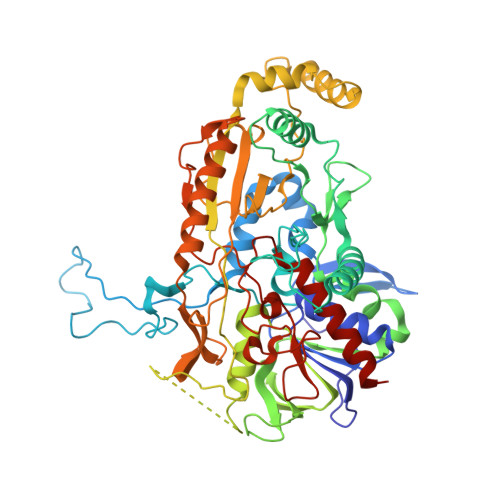
    The flavin-dependent homotetrameric enzyme pyranose 2-oxidase (P2O) is found mostly, but not exclusively, in lignocellulose-degrading fungi where it catalyzes the oxidation of β-d-glucose to the corresponding 2-keto sugar concomitantly with hydrogen peroxide formation during lignin solubilization. Here, we present crystal structures of P2O from the efficient lignocellulolytic basidiomycete Phanerochaete chrysosporium. Structures were determined of wild-type PcP2O from the natural fungal source, and two variants of recombinant full-length PcP2O, both in complex with the slow substrate 3-deoxy-3-fluoro-β-d-glucose. The active sites in PcP2O and P2O from Trametes multicolor (TmP2O) are highly conserved with identical substrate binding. Our structural analysis suggests that the 17 °C higher melting temperature of PcP2O compared to TmP2O is due to an increased number of intersubunit salt bridges. The structure of recombinant PcP2O expressed with its natural N-terminal sequence, including a proposed propeptide segment, reveals that the first five residues of the propeptide intercalate at the interface between A and B subunits to form stabilizing, mainly hydrophobic, interactions. In the structure of mature PcP2O purified from the natural source, the propeptide segment in subunit A has been replaced by a nearby loop in the B subunit. We propose that the propeptide in subunit A stabilizes the A/B interface of essential dimers in the homotetramer and that, upon maturation, it is replaced by the loop in the B subunit to form the mature subunit interface. This would imply that the propeptide segment of PcP2O acts as an intramolecular chaperone for oligomerization at the A/B interface of the essential dimer.


  • Organizational Affiliation

    KTH Royal Institute of Technology, School of Biotechnology, Albanova University Center, Roslagstullsbacken 21, S-10691 Stockholm Sweden.


Macromolecules
Find similar proteins by:  (by identity cutoff)  |  3D Structure
Entity ID: 1
MoleculeChains Sequence LengthOrganismDetailsImage
Pyranose 2-oxidase
A, B, C, D, E
A, B, C, D, E, F, G, H
621Phanerodontia chrysosporiumMutation(s): 1 
Gene Names: p2op2oxpox
EC: 1.1.3.10
UniProt
Find proteins for Q6QWR1 (Phanerodontia chrysosporium)
Explore Q6QWR1 
Go to UniProtKB:  Q6QWR1
Entity Groups  
Sequence Clusters30% Identity50% Identity70% Identity90% Identity95% Identity100% Identity
UniProt GroupQ6QWR1
Sequence Annotations
Expand
  • Reference Sequence
Small Molecules
Ligands 2 Unique
IDChains Name / Formula / InChI Key2D Diagram3D Interactions
FAD
Query on FAD

Download Ideal Coordinates CCD File 
I [auth A]
K [auth B]
M [auth C]
O [auth D]
Q [auth E]
I [auth A],
K [auth B],
M [auth C],
O [auth D],
Q [auth E],
S [auth F],
U [auth G],
W [auth H]
FLAVIN-ADENINE DINUCLEOTIDE
C27 H33 N9 O15 P2
VWWQXMAJTJZDQX-UYBVJOGSSA-N
G3F
Query on G3F

Download Ideal Coordinates CCD File 
J [auth A]
L [auth B]
N [auth C]
P [auth D]
R [auth E]
J [auth A],
L [auth B],
N [auth C],
P [auth D],
R [auth E],
T [auth F],
V [auth G],
X [auth H]
3-deoxy-3-fluoro-beta-D-glucopyranose
C6 H11 F O5
BUMRBAMACDBPKO-AIECOIEWSA-N
Experimental Data & Validation

Experimental Data

  • Method: X-RAY DIFFRACTION
  • Resolution: 2.40 Å
  • R-Value Free: 0.265 
  • R-Value Work: 0.205 
  • R-Value Observed: 0.206 
  • Space Group: P 1 21 1
Unit Cell:
Length ( Å )Angle ( ˚ )
a = 110.1α = 90
b = 167.099β = 93.98
c = 168.472γ = 90
Software Package:
Software NamePurpose
MAR345data collection
PHASERphasing
PHENIXrefinement
XDSdata reduction
XSCALEdata scaling

Structure Validation

View Full Validation Report


Ligand Structure Quality Assessment 


Entry History 

Deposition Data

Revision History  (Full details and data files)

  • Version 1.0: 2013-12-11
    Type: Initial release
  • Version 1.1: 2017-11-15
    Changes: Data collection, Refinement description
  • Version 1.2: 2020-07-29
    Type: Remediation
    Reason: Carbohydrate remediation
    Changes: Data collection, Database references, Derived calculations
  • Version 1.3: 2024-02-28
    Changes: Data collection, Database references, Structure summary