4MCT

P. vulgaris HIGBA structure, crystal form 1


Experimental Data Snapshot

  • Method: X-RAY DIFFRACTION
  • Resolution: 2.80 Å
  • R-Value Free: 0.238 
  • R-Value Work: 0.197 
  • R-Value Observed: 0.201 

wwPDB Validation   3D Report Full Report


This is version 1.2 of the entry. See complete history


Literature

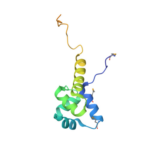
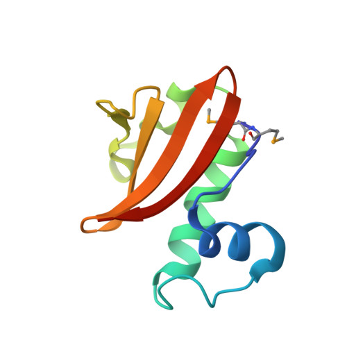
Structure of the Proteus vulgaris HigB-(HigA)2-HigB Toxin-Antitoxin Complex.

Schureck, M.A.Maehigashi, T.Miles, S.J.Marquez, J.Cho, S.E.Erdman, R.Dunham, C.M.

(2014) J Biol Chem 289: 1060-1070

  • DOI: https://doi.org/10.1074/jbc.M113.512095
  • Primary Citation of Related Structures:  
    4MCT, 4MCX

  • PubMed Abstract: 

    Bacterial toxin-antitoxin (TA) systems regulate key cellular processes to promote cell survival during periods of stress. During steady-state cell growth, antitoxins typically interact with their cognate toxins to inhibit activity presumably by preventing substrate recognition. We solved two x-ray crystal structures of the Proteus vulgaris tetrameric HigB-(HigA)2-HigB TA complex and found that, unlike most other TA systems, the antitoxin HigA makes minimal interactions with toxin HigB. HigB adopts a RelE family tertiary fold containing a highly conserved concave surface where we predict its active site is located. HigA does not cover the solvent-exposed HigB active site, suggesting that, in general, toxin inhibition is not solely mediated by active site hindrance by its antitoxin. Each HigA monomer contains a helix-turn-helix motif that binds to its own DNA operator to repress transcription during normal cellular growth. This is distinct from antitoxins belonging to other superfamilies that typically only form DNA-binding motifs upon dimerization. We further show that disruption of the HigB-(HigA)2-HigB tetramer to a HigBA heterodimer ablates operator binding. Taken together, our biochemical and structural studies elucidate the novel molecular details of the HigBA TA system.


  • Organizational Affiliation

    From the Department of Biochemistry, Emory University School of Medicine, Atlanta, Georgia 30322.


Macromolecules
Find similar proteins by:  (by identity cutoff)  |  3D Structure
Entity ID: 1
MoleculeChains Sequence LengthOrganismDetailsImage
Antidote protein
A, C
125Proteus vulgarisMutation(s): 0 
Gene Names: higA
UniProt
Find proteins for Q7A224 (Proteus vulgaris)
Explore Q7A224 
Go to UniProtKB:  Q7A224
Entity Groups  
Sequence Clusters30% Identity50% Identity70% Identity90% Identity95% Identity100% Identity
UniProt GroupQ7A224
Sequence Annotations
Expand
  • Reference Sequence
Find similar proteins by:  (by identity cutoff)  |  3D Structure
Entity ID: 2
MoleculeChains Sequence LengthOrganismDetailsImage
Killer protein
B, D
93Proteus vulgarisMutation(s): 0 
Gene Names: higB
UniProt
Find proteins for Q7A225 (Proteus vulgaris)
Explore Q7A225 
Go to UniProtKB:  Q7A225
Entity Groups  
Sequence Clusters30% Identity50% Identity70% Identity90% Identity95% Identity100% Identity
UniProt GroupQ7A225
Sequence Annotations
Expand
  • Reference Sequence
Small Molecules
Modified Residues  1 Unique
IDChains TypeFormula2D DiagramParent
MSE
Query on MSE
A, C
L-PEPTIDE LINKINGC5 H11 N O2 SeMET
Experimental Data & Validation

Experimental Data

  • Method: X-RAY DIFFRACTION
  • Resolution: 2.80 Å
  • R-Value Free: 0.238 
  • R-Value Work: 0.197 
  • R-Value Observed: 0.201 
  • Space Group: P 32 2 1
Unit Cell:
Length ( Å )Angle ( ˚ )
a = 94.86α = 90
b = 94.86β = 90
c = 126.814γ = 120
Software Package:
Software NamePurpose
DENZOdata reduction
SCALEPACKdata scaling
PHENIXrefinement
PDB_EXTRACTdata extraction
HKL-2000data reduction
HKL-2000data scaling
AutoSolphasing

Structure Validation

View Full Validation Report


Entry History 

Deposition Data

Revision History  (Full details and data files)

  • Version 1.0: 2013-12-11
    Type: Initial release
  • Version 1.1: 2014-01-29
    Changes: Database references
  • Version 1.2: 2017-11-15
    Changes: Refinement description