Design of reversible, cysteine-targeted Michael acceptors guided by kinetic and computational analysis.
Krishnan, S., Miller, R.M., Tian, B., Mullins, R.D., Jacobson, M.P., Taunton, J.(2014) J Am Chem Soc 136: 12624-12630
- PubMed: 25153195 Search on PubMedSearch on PubMed Central
- DOI: https://doi.org/10.1021/ja505194w
- Primary Citation Related Structures:
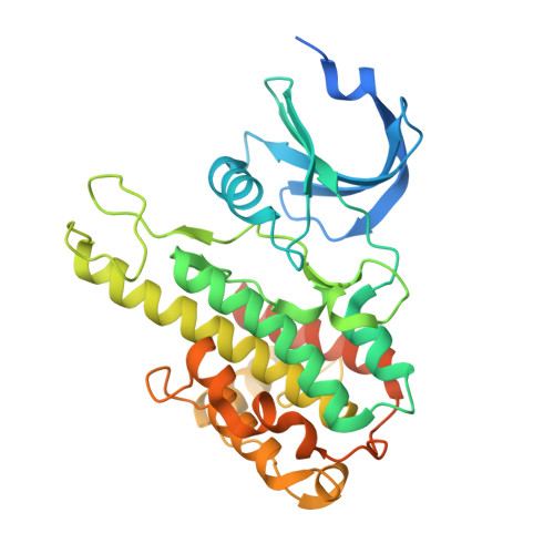
4MAO - PubMed Abstract:
Electrophilic probes that covalently modify a cysteine thiol often show enhanced pharmacological potency and selectivity. Although reversible Michael acceptors have been reported, the structural requirements for reversibility are poorly understood. Here, we report a novel class of acrylonitrile-based Michael acceptors, activated by aryl or heteroaryl electron-withdrawing groups. We demonstrate that thiol adducts of these acrylonitriles undergo β-elimination at rates that span more than 3 orders of magnitude. These rates correlate inversely with the computed proton affinity of the corresponding carbanions, enabling the intrinsic reversibility of the thiol-Michael reaction to be tuned in a predictable manner. We apply these principles to the design of new reversible covalent kinase inhibitors with improved properties. A cocrystal structure of one such inhibitor reveals specific noncovalent interactions between the 1,2,4-triazole activating group and the kinase. Our experimental and computational study enables the design of new Michael acceptors, expanding the palette of reversible, cysteine-targeted electrophiles.
- Department of Cellular and Molecular Pharmacology, Howard Hughes Medical Institute, University of California-San Francisco , San Francisco, California 94158, United States.
Organizational Affiliation: