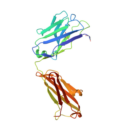
4M93

Unliganded 2 crystal structure of S25-26 Fab


Experimental Data Snapshot

  • Method: X-RAY DIFFRACTION
  • Resolution: 2.09 Å
  • R-Value Free: 0.251 
  • R-Value Work: 0.218 
  • R-Value Observed: 0.219 

wwPDB Validation   3D Report Full Report


Ligand Structure Quality Assessment 


This is version 2.1 of the entry. See complete history


Literature

Groove-type Recognition of Chlamydiaceae-specific Lipopolysaccharide Antigen by a Family of Antibodies Possessing an Unusual Variable Heavy Chain N-Linked Glycan.

Haji-Ghassemi, O.Muller-Loennies, S.Saldova, R.Muniyappa, M.Brade, L.Rudd, P.M.Harvey, D.J.Kosma, P.Brade, H.Evans, S.V.

(2014) J Biol Chem 289: 16644-16661

  • DOI: https://doi.org/10.1074/jbc.M113.528224
  • Primary Citation of Related Structures:  
    4M7J, 4M7Z, 4M93, 4MA1

  • PubMed Abstract: 
  • The structure of the antigen binding fragment of mAb S25-26, determined to 1.95 Å resolution in complex with the Chlamydiaceae family-specific trisaccharide antigen Kdo(2→8)Kdo(2→4)Kdo (Kdo = 3-deoxy-α-d-manno-oct-2-ulopyranosonic acid), displays a germ-line-coded paratope that differs significantly from previously characterized Chlamydiaceae-specific mAbs despite being raised against the identical immunogen ...

    The structure of the antigen binding fragment of mAb S25-26, determined to 1.95 Å resolution in complex with the Chlamydiaceae family-specific trisaccharide antigen Kdo(2→8)Kdo(2→4)Kdo (Kdo = 3-deoxy-α-d-manno-oct-2-ulopyranosonic acid), displays a germ-line-coded paratope that differs significantly from previously characterized Chlamydiaceae-specific mAbs despite being raised against the identical immunogen. Unlike the terminal Kdo recognition pocket that promotes cross-reactivity in S25-2-type antibodies, S25-26 and the closely related S25-23 utilize a groove composed of germ-line residues to recognize the entire trisaccharide antigen and so confer strict specificity. Interest in S25-23 was sparked by its rare high μm affinity and strict specificity for the family-specific trisaccharide antigen; however, only the related antibody S25-26 proved amenable to crystallization. The structures of three unliganded forms of S25-26 have a labile complementary-determining region H3 adjacent to significant glycosylation of the variable heavy chain on asparagine 85 in Framework Region 3. Analysis of the glycan reveals a heterogeneous mixture with a common root structure that contains an unusually high number of terminal αGal-Gal moieties. One of the few reported structures of glycosylated mAbs containing these epitopes is the therapeutic antibody Cetuximab; however, unlike Cetuximab, one of the unliganded structures in S25-26 shows significant order in the glycan with appropriate electron density for nine residues. The elucidation of the three-dimensional structure of an αGal-containing N-linked glycan on a mAb variable heavy chain has potential clinical interest, as it has been implicated in allergic response in patients receiving therapeutic antibodies.


    Organizational Affiliation

    From the Department of Biochemistry and Microbiology, University of Victoria, Victoria, British Columbia V8P 3P6, Canada, svevans@uvic.ca.


Macromolecules
Find similar proteins by:  (by identity cutoff)  |  3D Structure
Entity ID: 1
MoleculeChainsSequence LengthOrganismDetailsImage
S25-26 Fab (IgG1k) light chainA [auth L],
C
219Mus musculusMutation(s): 0 
Entity Groups  
Sequence Clusters30% Identity50% Identity70% Identity90% Identity95% Identity100% Identity
Protein Feature View
Expand
  • Reference Sequence
Find similar proteins by:  (by identity cutoff)  |  3D Structure
Entity ID: 2
MoleculeChainsSequence LengthOrganismDetailsImage
S25-26 Fab (IgG1k) heavy chainB [auth H],
D [auth B]
218Mus musculusMutation(s): 0 
Entity Groups  
Sequence Clusters30% Identity50% Identity70% Identity90% Identity95% Identity100% Identity
Protein Feature View
Expand
  • Reference Sequence
Small Molecules
Ligands 3 Unique
IDChainsName / Formula / InChI Key2D Diagram3D Interactions
NAG
Query on NAG

Download Ideal Coordinates CCD File 
E [auth H],
F [auth B]
2-acetamido-2-deoxy-beta-D-glucopyranose
C8 H15 N O6
OVRNDRQMDRJTHS-FMDGEEDCSA-N
 Ligand Interaction
ACT
Query on ACT

Download Ideal Coordinates CCD File 
H [auth B],
I [auth B],
J [auth B]
ACETATE ION
C2 H3 O2
QTBSBXVTEAMEQO-UHFFFAOYSA-M
 Ligand Interaction
CA
Query on CA

Download Ideal Coordinates CCD File 
G [auth B]CALCIUM ION
Ca
BHPQYMZQTOCNFJ-UHFFFAOYSA-N
 Ligand Interaction
Modified Residues  1 Unique
IDChainsTypeFormula2D DiagramParent
PCA
Query on PCA
B [auth H],
D [auth B]
L-PEPTIDE LINKINGC5 H7 N O3GLN
Experimental Data & Validation

Experimental Data

  • Method: X-RAY DIFFRACTION
  • Resolution: 2.09 Å
  • R-Value Free: 0.251 
  • R-Value Work: 0.218 
  • R-Value Observed: 0.219 
  • Space Group: C 1 2 1
Unit Cell:
Length ( Å )Angle ( ˚ )
a = 118.894α = 90
b = 88.806β = 102.06
c = 101.89γ = 90
Software Package:
Software NamePurpose
DENZOdata reduction
SCALEPACKdata scaling
PHASERphasing
REFMACrefinement
PDB_EXTRACTdata extraction
Macromoleculardata collection

Structure Validation

View Full Validation Report


Ligand Structure Quality Assessment 


Entry History 

Deposition Data

Revision History  (Full details and data files)

  • Version 1.0: 2014-04-09
    Type: Initial release
  • Version 1.1: 2014-06-11
    Changes: Database references
  • Version 1.2: 2014-07-09
    Changes: Database references
  • Version 1.3: 2017-11-15
    Changes: Refinement description
  • Version 2.0: 2019-12-25
    Changes: Derived calculations, Polymer sequence
  • Version 2.1: 2020-07-29
    Type: Remediation
    Reason: Carbohydrate remediation
    Changes: Data collection, Derived calculations, Structure summary