4M8D

Crystal structure of an isatin hydrolase bound to product analogue thioisatinate


Experimental Data Snapshot

  • Method: X-RAY DIFFRACTION
  • Resolution: 1.90 Å
  • R-Value Free: 0.192 
  • R-Value Work: 0.152 
  • R-Value Observed: 0.154 

wwPDB Validation   3D Report Full Report


Ligand Structure Quality Assessment 


This is version 1.4 of the entry. See complete history


Literature

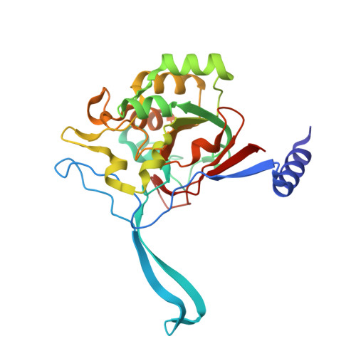
A proton wire and water channel revealed in the crystal structure of isatin hydrolase.

Bjerregaard-Andersen, K.Sommer, T.Jensen, J.K.Jochimsen, B.Etzerodt, M.Morth, J.P.

(2014) J Biol Chem 289: 21351-21359

  • DOI: https://doi.org/10.1074/jbc.M114.568824
  • Primary Citation of Related Structures:  
    4J0N, 4M8D

  • PubMed Abstract: 

    The high resolution crystal structures of isatin hydrolase from Labrenzia aggregata in the apo and the product state are described. These are the first structures of a functionally characterized metal-dependent hydrolase of this fold. Isatin hydrolase converts isatin to isatinate and belongs to a novel family of metalloenzymes that include the bacterial kynurenine formamidase. The product state, mimicked by bound thioisatinate, reveals a water molecule that bridges the thioisatinate to a proton wire in an adjacent water channel and thus allows the proton released by the reaction to escape only when the product is formed. The functional proton wire present in isatin hydrolase isoform b represents a unique catalytic feature common to all hydrolases is here trapped and visualized for the first time. The local molecular environment required to coordinate thioisatinate allows stronger and more confident identification of orthologous genes encoding isatin hydrolases within the prokaryotic kingdom. The isatin hydrolase orthologues found in human gut bacteria raise the question as to whether the indole-3-acetic acid degradation pathway is present in human gut flora.


  • Organizational Affiliation

    From the Norwegian Center of Molecular Medicine, Nordic EMBL Partnership University of Oslo, Gaustadalléen 21, 0349 Oslo, Norway, the Department for Molecular Biology and Genetics, Aarhus University, Gustav Wieds vej 10C, DK-8000 Aarhus, Denmark.


Macromolecules
Find similar proteins by:  (by identity cutoff)  |  3D Structure
Entity ID: 1
MoleculeChains Sequence LengthOrganismDetailsImage
Putative uncharacterized protein
A, B, C, D, E
A, B, C, D, E, F, G, H, I, J, K, L
263Roseibium aggregatum IAM 12614Mutation(s): 0 
Gene Names: SIAM614_09648
UniProt
Find proteins for A0NLY7 (Roseibium aggregatum (strain ATCC 25650 / DSM 13394 / JCM 20685 / NBRC 16684 / NCIMB 2208 / IAM 12614 / B1))
Explore A0NLY7 
Go to UniProtKB:  A0NLY7
Entity Groups  
Sequence Clusters30% Identity50% Identity70% Identity90% Identity95% Identity100% Identity
UniProt GroupA0NLY7
Sequence Annotations
Expand
  • Reference Sequence
Small Molecules
Ligands 3 Unique
IDChains Name / Formula / InChI Key2D Diagram3D Interactions
23J
Query on 23J

Download Ideal Coordinates CCD File 
BA [auth D]
EA [auth E]
GA [auth F]
JA [auth G]
LA [auth H]
BA [auth D],
EA [auth E],
GA [auth F],
JA [auth G],
LA [auth H],
N [auth A],
NA [auth I],
QA [auth J],
S [auth B],
UA [auth K],
WA [auth L],
X [auth C]
oxo(2-sulfanylphenyl)acetic acid
C8 H6 O3 S
QAZZLGCIPWJVRJ-UHFFFAOYSA-N
MN
Query on MN

Download Ideal Coordinates CCD File 
AA [auth D]
DA [auth E]
FA [auth F]
IA [auth G]
KA [auth H]
AA [auth D],
DA [auth E],
FA [auth F],
IA [auth G],
KA [auth H],
M [auth A],
MA [auth I],
PA [auth J],
R [auth B],
TA [auth K],
VA [auth L],
W [auth C]
MANGANESE (II) ION
Mn
WAEMQWOKJMHJLA-UHFFFAOYSA-N
CA
Query on CA

Download Ideal Coordinates CCD File 
CA [auth D]
HA [auth F]
O [auth A]
OA [auth I]
P [auth A]
CA [auth D],
HA [auth F],
O [auth A],
OA [auth I],
P [auth A],
Q [auth A],
RA [auth J],
SA [auth J],
T [auth B],
U [auth B],
V [auth B],
XA [auth L],
Y [auth C],
YA [auth L],
Z [auth C],
ZA [auth L]
CALCIUM ION
Ca
BHPQYMZQTOCNFJ-UHFFFAOYSA-N
Binding Affinity Annotations 
IDSourceBinding Affinity
23J Binding MOAD:  4M8D IC50: 1.00e+4 (nM) from 1 assay(s)
Experimental Data & Validation

Experimental Data

  • Method: X-RAY DIFFRACTION
  • Resolution: 1.90 Å
  • R-Value Free: 0.192 
  • R-Value Work: 0.152 
  • R-Value Observed: 0.154 
  • Space Group: P 1 21 1
Unit Cell:
Length ( Å )Angle ( ˚ )
a = 84.98α = 90
b = 51.82β = 91.92
c = 357.09γ = 90
Software Package:
Software NamePurpose
MxCuBEdata collection
PHENIXmodel building
PHENIXrefinement
XDSdata reduction
XSCALEdata scaling
PHENIXphasing

Structure Validation

View Full Validation Report


Ligand Structure Quality Assessment 


Entry History 

Deposition Data

Revision History  (Full details and data files)

  • Version 1.0: 2014-06-18
    Type: Initial release
  • Version 1.1: 2014-07-16
    Changes: Database references
  • Version 1.2: 2014-08-20
    Changes: Database references
  • Version 1.3: 2018-03-07
    Changes: Data collection
  • Version 1.4: 2023-09-20
    Changes: Data collection, Database references, Derived calculations, Refinement description