4M8B

Fungal Protein


Experimental Data Snapshot

  • Method: X-RAY DIFFRACTION
  • Resolution: 2.61 Å
  • R-Value Free: 0.235 
  • R-Value Work: 0.198 

wwPDB Validation   3D Report Full Report


This is version 1.5 of the entry. See complete history


Literature

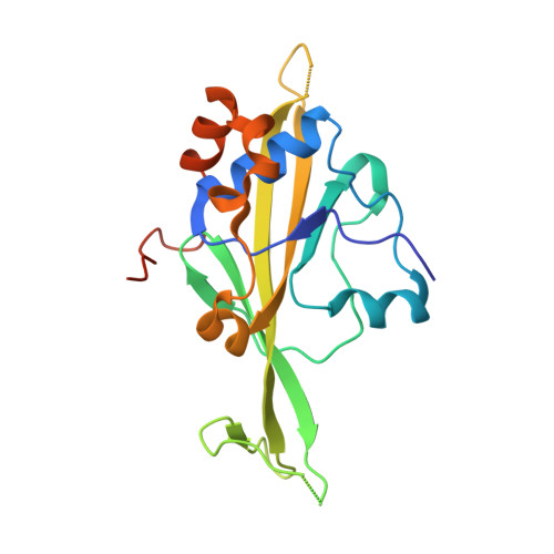
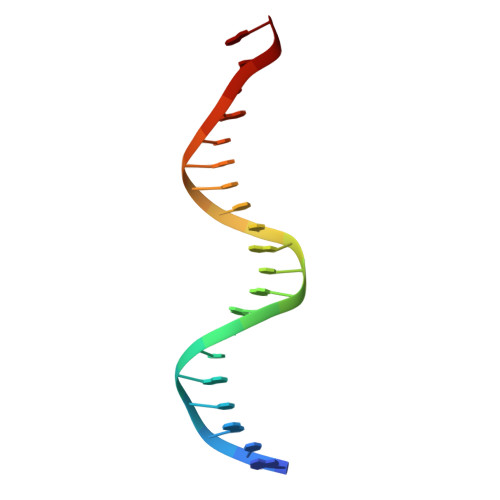
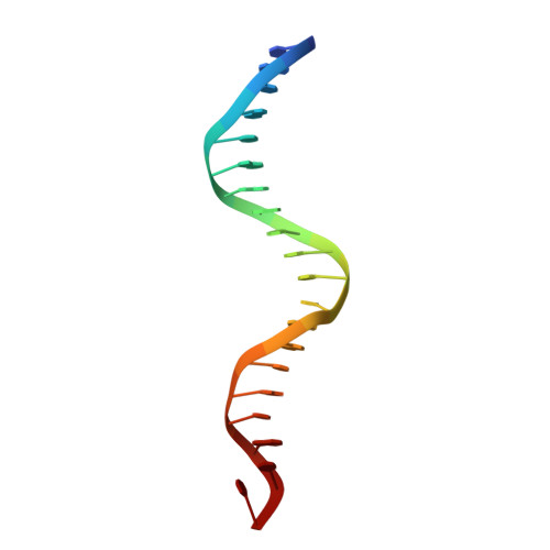
Structure of a new DNA-binding domain which regulates pathogenesis in a wide variety of fungi.

Lohse, M.B.Rosenberg, O.S.Cox, J.S.Stroud, R.M.Finer-Moore, J.S.Johnson, A.D.

(2014) Proc Natl Acad Sci U S A 111: 10404-10410

  • DOI: https://doi.org/10.1073/pnas.1410110111
  • Primary Citation of Related Structures:  
    4M8B

  • PubMed Abstract: 

    WOPR-domain proteins are found throughout the fungal kingdom where they function as master regulators of cell morphology and pathogenesis. Genetic and biochemical experiments previously demonstrated that these proteins bind to specific DNA sequences and thereby regulate transcription. However, their primary sequence showed no relationship to any known DNA-binding domain, and the basis for their ability to recognize DNA sequences remained unknown. Here, we describe the 2.6-Å crystal structure of a WOPR domain in complex with its preferred DNA sequence. The structure reveals that two highly conserved regions, separated by an unconserved linker, form an interdigitated β-sheet that is tilted into the major groove of DNA. Although the main interaction surface is in the major groove, the highest-affinity interactions occur in the minor groove, primarily through a deeply penetrating arginine residue. The structure reveals a new, unanticipated mechanism by which proteins can recognize specific sequences of DNA.


  • Organizational Affiliation

    Departments of Microbiology and Immunology and.


Macromolecules

Find similar proteins by:  (by identity cutoff)  |  3D Structure
Entity ID: 3
MoleculeChains Sequence LengthOrganismDetailsImage
YHR177WC [auth R]202Saccharomyces cerevisiae S288CMutation(s): 0 
Gene Names: YHR177WYhr177wp
UniProt
Find proteins for P38867 (Saccharomyces cerevisiae (strain ATCC 204508 / S288c))
Explore P38867 
Go to UniProtKB:  P38867
Entity Groups  
Sequence Clusters30% Identity50% Identity70% Identity90% Identity95% Identity100% Identity
UniProt GroupP38867
Sequence Annotations
Expand
  • Reference Sequence

Find similar nucleic acids by:  Sequence   |   3D Structure  

Entity ID: 1
MoleculeChains LengthOrganismImage
Chain A of dsDNA containing the cis-regulatory element20N/A
Sequence Annotations
Expand
  • Reference Sequence

Find similar nucleic acids by:  Sequence   |   3D Structure  

Entity ID: 2
MoleculeChains LengthOrganismImage
Chain B of dsDNA containing the cis-regulatory element20N/A
Sequence Annotations
Expand
  • Reference Sequence
Small Molecules
Ligands 1 Unique
IDChains Name / Formula / InChI Key2D Diagram3D Interactions
EDO
Query on EDO

Download Ideal Coordinates CCD File 
D [auth R]1,2-ETHANEDIOL
C2 H6 O2
LYCAIKOWRPUZTN-UHFFFAOYSA-N
Experimental Data & Validation

Experimental Data

  • Method: X-RAY DIFFRACTION
  • Resolution: 2.61 Å
  • R-Value Free: 0.235 
  • R-Value Work: 0.198 
  • Space Group: P 21 21 21
Unit Cell:
Length ( Å )Angle ( ˚ )
a = 90.293α = 90
b = 103.401β = 90
c = 73.178γ = 90
Software Package:
Software NamePurpose
ADSCdata collection
SOLVEphasing
PHENIXrefinement
MOSFLMdata reduction
SCALAdata scaling

Structure Validation

View Full Validation Report


Entry History 

Deposition Data

Revision History  (Full details and data files)

  • Version 1.0: 2014-06-25
    Type: Initial release
  • Version 1.1: 2014-07-02
    Changes: Database references
  • Version 1.2: 2014-07-23
    Changes: Database references
  • Version 1.3: 2014-08-20
    Changes: Database references
  • Version 1.4: 2024-02-28
    Changes: Data collection, Database references, Derived calculations
  • Version 1.5: 2024-04-03
    Changes: Refinement description