The Lid Domain of the MCP Hydrolase DxnB2 Contributes to the Reactivity toward Recalcitrant PCB Metabolites.
Ruzzini, A.C., Bhowmik, S., Yam, K.C., Ghosh, S., Bolin, J.T., Eltis, L.D.(2013) Biochemistry 52: 5685-5695
- PubMed: 23879719 Search on PubMedSearch on PubMed Central
- DOI: https://doi.org/10.1021/bi400774m
- Primary Citation Related Structures:
4LXG, 4LXH, 4LYD - PubMed Abstract:
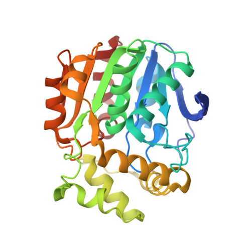
DxnB2 and BphD are meta-cleavage product (MCP) hydrolases that catalyze C-C bond hydrolysis of the biphenyl metabolite 2-hydroxy-6-oxo-6-phenylhexa-2,4-dienoic acid (HOPDA). BphD is a bottleneck in the bacterial degradation of polychlorinated biphenyls (PCBs) by the Bph catabolic pathway due in part to inhibition by 3-Cl HOPDAs. By contrast, DxnB2 from Sphingomonas wittichii RW1 catalyzes the hydrolysis of 3-Cl HOPDAs more efficiently. X-ray crystallographic studies of the catalytically inactive S105A variant of DxnB2 complexed with 3-Cl HOPDA revealed a binding mode in which C1 through C6 of the dienoate are coplanar. The chlorine substituent is accommodated by a hydrophobic pocket that is larger than the homologous site in BphDLB400 from Burkholderia xenovorans LB400. The planar binding mode observed in the crystalline complex was consistent with the hyper- and hypsochromically shifted absorption spectra of 3-Cl and 3,9,11-triCl HOPDA, respectively, bound to S105A in solution. Moreover, ES(red), an intermediate possessing a bathochromically shifted spectrum observed in the turnover of HOPDA, was not detected, suggesting that substrate destabilization was rate-limiting in the turnover of these PCB metabolites. Interestingly, electron density for the first α-helix of the lid domain was poorly defined in the dimeric DxnB2 structures, unlike in the tetrameric BphDLB400. Structural comparison of MCP hydrolases identified the NC-loop, connecting the lid to the α/β-hydrolase core domain, as a determinant in the oligomeric state and suggests its involvement in catalysis. Finally, an increased mobility of the DxnB2 lid may contribute to the enzyme's ability to hydrolyze PCB metabolites, highlighting how lid architecture contributes to substrate specificity in α/β-hydrolases.
- Department of Biochemistry and Molecular Biology, University of British Columbia, BC, Canada.
Organizational Affiliation: