


Crystal structure of a glycosomal glyceraldehyde-3-phosphate dehydrogenase from Trypanosoma cruzi
Edwards, T.E., Abendroth, J.To be published.
Experimental Data Snapshot
Starting Model: experimental
View more details
Entity ID: 1 | |||||
|---|---|---|---|---|---|
| Molecule | Chains | Sequence Length | Organism | Details | Image |
| Glyceraldehyde 3-phosphate dehydrogenase, cytosolic | 346 | Trypanosoma cruzi strain CL Brener | Mutation(s): 0 Gene Names: Tc00.1047053510105.230 EC: 1.2.1.12 | 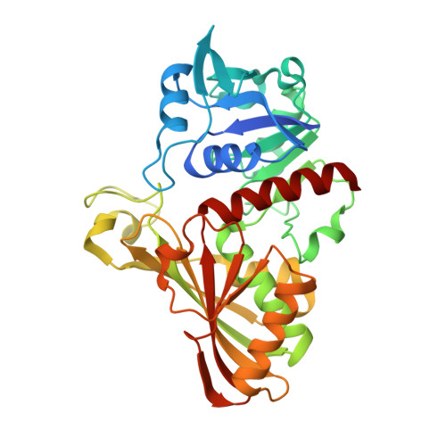 | |
UniProt | |||||
Entity Groups | |||||
| Sequence Clusters | 30% Identity50% Identity70% Identity90% Identity95% Identity100% Identity | ||||
| UniProt Group | Q4DZT1 | ||||
Sequence AnnotationsExpand | |||||
Reference Sequence | |||||
| Ligands 2 Unique | |||||
|---|---|---|---|---|---|
| ID | Chains | Name / Formula / InChI Key | 2D Diagram | 3D Interactions | |
| NAD Download:Ideal Coordinates CCD File | C [auth A], H [auth B] | NICOTINAMIDE-ADENINE-DINUCLEOTIDE C21 H27 N7 O14 P2 BAWFJGJZGIEFAR-NNYOXOHSSA-N |  | ||
| EDO Download:Ideal Coordinates CCD File | D [auth A] E [auth A] F [auth A] G [auth A] I [auth B] | 1,2-ETHANEDIOL C2 H6 O2 LYCAIKOWRPUZTN-UHFFFAOYSA-N |  | ||
| Length ( Å ) | Angle ( ˚ ) |
|---|---|
| a = 80.49 | α = 90 |
| b = 80.49 | β = 90 |
| c = 203.06 | γ = 120 |
| Software Name | Purpose |
|---|---|
| XSCALE | data scaling |
| PHASER | phasing |
| REFMAC | refinement |
| PDB_EXTRACT | data extraction |
| XDS | data reduction |


