4LNC

Neutron structure of the cyclic glucose bound Xylose Isomerase E186Q mutant


Experimental Data Snapshot

  • Method: NEUTRON DIFFRACTION
  • Resolution: 2.19 Å
  • R-Value Free: 0.322 
  • R-Value Work: 0.284 
  • R-Value Observed: 0.288 

wwPDB Validation   3D Report Full Report


This is version 1.5 of the entry. See complete history


Literature

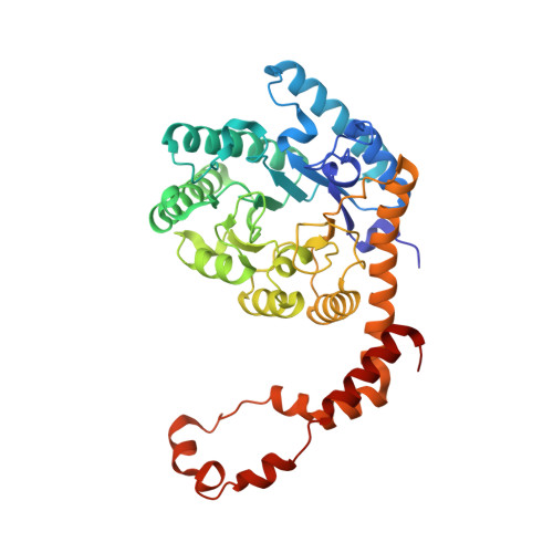
Neutron structure of the cyclic glucose-bound xylose isomerase E186Q mutant.

Munshi, P.Snell, E.H.van der Woerd, M.J.Judge, R.A.Myles, D.A.Ren, Z.Meilleur, F.

(2014) Acta Crystallogr D Biol Crystallogr 70: 414-420

  • DOI: https://doi.org/10.1107/S1399004713029684
  • Primary Citation of Related Structures:  
    4LNC

  • PubMed Abstract: 

    Ketol-isomerases catalyze the reversible isomerization between aldoses and ketoses. D-Xylose isomerase carries out the first reaction in the catabolism of D-xylose, but is also able to convert D-glucose to D-fructose. The first step of the reaction is an enzyme-catalyzed ring opening of the cyclic substrate. The active-site amino-acid acid/base pair involved in ring opening has long been investigated and several models have been proposed. Here, the structure of the xylose isomerase E186Q mutant with cyclic glucose bound at the active site, refined against joint X-ray and neutron diffraction data, is reported. Detailed analysis of the hydrogen-bond networks at the active site of the enzyme suggests that His54, which is doubly protonated, is poised to protonate the glucose O5 position, while Lys289, which is neutral, promotes deprotonation of the glucose O1H hydroxyl group via an activated water molecule. The structure also reveals an extended hydrogen-bonding network that connects the conserved residues Lys289 and Lys183 through three structurally conserved water molecules and residue 186, which is a glutamic acid to glutamine mutation.


  • Organizational Affiliation

    Neutron Sciences Directorate, Oak Ridge National Laboratory, Oak Ridge, TN 37831, USA.


Macromolecules
Find similar proteins by:  (by identity cutoff)  |  3D Structure
Entity ID: 1
MoleculeChains Sequence LengthOrganismDetailsImage
Xylose isomerase388Streptomyces rubiginosusMutation(s): 3 
Gene Names: xylA
EC: 5.3.1.5
UniProt
Find proteins for P24300 (Streptomyces rubiginosus)
Explore P24300 
Go to UniProtKB:  P24300
Entity Groups  
Sequence Clusters30% Identity50% Identity70% Identity90% Identity95% Identity100% Identity
UniProt GroupP24300
Sequence Annotations
Expand
  • Reference Sequence
Small Molecules
Ligands 3 Unique
IDChains Name / Formula / InChI Key2D Diagram3D Interactions
GLC
Query on GLC

Download Ideal Coordinates CCD File 
B [auth A]alpha-D-glucopyranose
C6 H12 O6
WQZGKKKJIJFFOK-DVKNGEFBSA-N
MN
Query on MN

Download Ideal Coordinates CCD File 
C [auth A],
D [auth A]
MANGANESE (II) ION
Mn
WAEMQWOKJMHJLA-UHFFFAOYSA-N
MG
Query on MG

Download Ideal Coordinates CCD File 
E [auth A]MAGNESIUM ION
Mg
JLVVSXFLKOJNIY-UHFFFAOYSA-N
Experimental Data & Validation

Experimental Data

  • Method: NEUTRON DIFFRACTION
  • Resolution: 2.19 Å
  • R-Value Free: 0.322 
  • R-Value Work: 0.284 
  • R-Value Observed: 0.288 
  • Space Group: I 2 2 2
Unit Cell:
Length ( Å )Angle ( ˚ )
a = 93.97α = 90
b = 99.52β = 90
c = 102.92γ = 90
Software Package:
Software NamePurpose
Quasi-Lauedata collection
PHENIXrefinement
LAUEGENdata reduction
SCALAdata scaling
PHENIXphasing

Structure Validation

View Full Validation Report


Entry History 

Deposition Data

Revision History  (Full details and data files)

  • Version 1.0: 2014-02-12
    Type: Initial release
  • Version 1.1: 2014-09-24
    Changes: Database references
  • Version 1.2: 2018-04-25
    Changes: Data collection
  • Version 1.3: 2018-06-13
    Changes: Data collection, Experimental preparation
  • Version 1.4: 2020-07-29
    Type: Remediation
    Reason: Carbohydrate remediation
    Changes: Data collection, Database references, Derived calculations, Structure summary
  • Version 1.5: 2024-02-28
    Changes: Data collection, Database references, Structure summary