4LNA

CRYSTAL STRUCTURE OF purine nucleoside phosphorylase I from Spirosoma linguale DSM 74, NYSGRC Target 029362


Experimental Data Snapshot

  • Method: X-RAY DIFFRACTION
  • Resolution: 2.10 Å
  • R-Value Free: 0.216 
  • R-Value Work: 0.172 
  • R-Value Observed: 0.174 

wwPDB Validation   3D Report Full Report


This is version 1.1 of the entry. See complete history


Literature

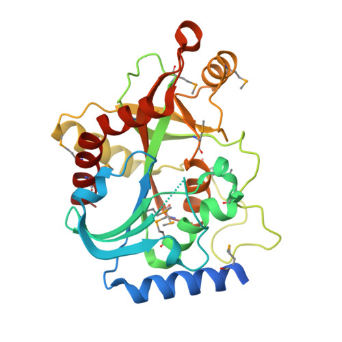
Crystal structure of purine nucleoside phosphorylase I from Spirosoma linguale DSM 74, NYSGRC Target 029362

Malashkevich, V.N.Bonanno, J.B.Bhosle, R.Toro, R.Hillerich, B.Gizzi, A.Garforth, S.Kar, A.Chan, M.K.Lafluer, J.Patel, H.Matikainen, B.Chamala, S.Lim, S.Celikgil, A.Villegas, G.Evans, B.Love, J.Fiser, A.Khafizov, K.Seidel, R.Almo, S.C.

To be published.

Macromolecules
Find similar proteins by:  (by identity cutoff)  |  3D Structure
Entity ID: 1
MoleculeChainsSequence LengthOrganismDetailsImage
Purine nucleoside phosphorylase293Spirosoma linguale DSM 74Mutation(s): 0 
Gene Names: 8727701Slin_3943
EC: 2.4.2.1
UniProt
Find proteins for D2QIK7 (Spirosoma linguale (strain ATCC 33905 / DSM 74 / LMG 10896 / Claus 1))
Explore D2QIK7 
Go to UniProtKB:  D2QIK7
Entity Groups  
Sequence Clusters30% Identity50% Identity70% Identity90% Identity95% Identity100% Identity
UniProt GroupD2QIK7
Protein Feature View
Expand
  • Reference Sequence
Small Molecules
Ligands 4 Unique
IDChainsName / Formula / InChI Key2D Diagram3D Interactions
ADE
Query on ADE

Download Ideal Coordinates CCD File 
D [auth A]ADENINE
C5 H5 N5
GFFGJBXGBJISGV-UHFFFAOYSA-N
 Ligand Interaction
MPD
Query on MPD

Download Ideal Coordinates CCD File 
E [auth A](4S)-2-METHYL-2,4-PENTANEDIOL
C6 H14 O2
SVTBMSDMJJWYQN-YFKPBYRVSA-N
 Ligand Interaction
PO4
Query on PO4

Download Ideal Coordinates CCD File 
B [auth A],
C [auth A]
PHOSPHATE ION
O4 P
NBIIXXVUZAFLBC-UHFFFAOYSA-K
 Ligand Interaction
CL
Query on CL

Download Ideal Coordinates CCD File 
F [auth A]CHLORIDE ION
Cl
VEXZGXHMUGYJMC-UHFFFAOYSA-M
 Ligand Interaction
Modified Residues  1 Unique
IDChainsTypeFormula2D DiagramParent
MSE
Query on MSE
A
L-PEPTIDE LINKINGC5 H11 N O2 SeMET
Experimental Data & Validation

Experimental Data

  • Method: X-RAY DIFFRACTION
  • Resolution: 2.10 Å
  • R-Value Free: 0.216 
  • R-Value Work: 0.172 
  • R-Value Observed: 0.174 
  • Space Group: P 43 3 2
Unit Cell:
Length ( Å )Angle ( ˚ )
a = 125.561α = 90
b = 125.561β = 90
c = 125.561γ = 90
Software Package:
Software NamePurpose
SCALEPACKdata scaling
PHASERphasing
REFMACrefinement
PDB_EXTRACTdata extraction
CBASSdata collection
HKL-2000data reduction

Structure Validation

View Full Validation Report


Entry History 

Revision History  (Full details and data files)

  • Version 1.0: 2013-07-24
    Type: Initial release
  • Version 1.1: 2023-09-20
    Changes: Data collection, Database references, Derived calculations, Refinement description