4LJQ

Crystal structure of the catalytic core of E3 ligase HOIP


Experimental Data Snapshot

  • Method: X-RAY DIFFRACTION
  • Resolution: 2.45 Å
  • R-Value Free: 0.243 
  • R-Value Work: 0.206 
  • R-Value Observed: 0.209 

wwPDB Validation   3D Report Full Report


This is version 1.1 of the entry. See complete history


Literature

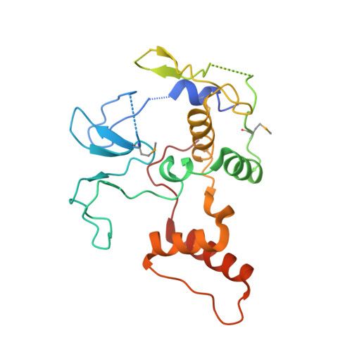
Structural basis for ligase-specific conjugation of linear ubiquitin chains by HOIP.

Stieglitz, B.Rana, R.R.Koliopoulos, M.G.Morris-Davies, A.C.Schaeffer, V.Christodoulou, E.Howell, S.Brown, N.R.Dikic, I.Rittinger, K.

(2013) Nature 503: 422-426

  • DOI: https://doi.org/10.1038/nature12638
  • Primary Citation of Related Structures:  
    4LJO, 4LJP, 4LJQ

  • PubMed Abstract: 

    Linear ubiquitin chains are important regulators of cellular signalling pathways that control innate immunity and inflammation through nuclear factor (NF)-κB activation and protection against tumour necrosis factor-α-induced apoptosis. They are synthesized by HOIP, which belongs to the RBR (RING-between-RING) family of E3 ligases and is the catalytic component of LUBAC (linear ubiquitin chain assembly complex), a multisubunit E3 ligase. RBR family members act as RING/HECT hybrids, employing RING1 to recognize ubiquitin-loaded E2 while a conserved cysteine in RING2 subsequently forms a thioester intermediate with the transferred or 'donor' ubiquitin. Here we report the crystal structure of the catalytic core of HOIP in its apo form and in complex with ubiquitin. The carboxy-terminal portion of HOIP adopts a novel fold that, together with a zinc-finger, forms a ubiquitin-binding platform that orients the acceptor ubiquitin and positions its α-amino group for nucleophilic attack on the E3∼ubiquitin thioester. The C-terminal tail of a second ubiquitin molecule is located in close proximity to the catalytic cysteine, providing a unique snapshot of the ubiquitin transfer complex containing both donor and acceptor ubiquitin. These interactions are required for activation of the NF-κB pathway in vivo, and they explain the determinants of linear ubiquitin chain specificity by LUBAC.


  • Organizational Affiliation

    Division of Molecular Structure, MRC-National Institute for Medical Research, The Ridgeway, London NW7 1AA, UK.


Macromolecules
Find similar proteins by:  (by identity cutoff)  |  3D Structure
Entity ID: 1
MoleculeChains Sequence LengthOrganismDetailsImage
E3 ubiquitin-protein ligase RNF31A [auth B],
B [auth A],
C,
D
223Homo sapiensMutation(s): 0 
Gene Names: RNF31ZIBRA
EC: 6.3.2
UniProt & NIH Common Fund Data Resources
Find proteins for Q96EP0 (Homo sapiens)
Explore Q96EP0 
Go to UniProtKB:  Q96EP0
PHAROS:  Q96EP0
GTEx:  ENSG00000092098 
Entity Groups  
Sequence Clusters30% Identity50% Identity70% Identity90% Identity95% Identity100% Identity
UniProt GroupQ96EP0
Sequence Annotations
Expand
  • Reference Sequence
Small Molecules
Ligands 1 Unique
IDChains Name / Formula / InChI Key2D Diagram3D Interactions
ZN
Query on ZN

Download Ideal Coordinates CCD File 
E [auth B]
F [auth B]
G [auth B]
H [auth B]
I [auth B]
E [auth B],
F [auth B],
G [auth B],
H [auth B],
I [auth B],
J [auth A],
K [auth A],
L [auth A],
M [auth A],
N [auth A],
O [auth C],
P [auth C],
Q [auth C],
R [auth C],
S [auth D],
T [auth D],
U [auth D],
V [auth D]
ZINC ION
Zn
PTFCDOFLOPIGGS-UHFFFAOYSA-N
Modified Residues  1 Unique
IDChains TypeFormula2D DiagramParent
MSE
Query on MSE
A [auth B],
B [auth A],
C,
D
L-PEPTIDE LINKINGC5 H11 N O2 SeMET
Experimental Data & Validation

Experimental Data

  • Method: X-RAY DIFFRACTION
  • Resolution: 2.45 Å
  • R-Value Free: 0.243 
  • R-Value Work: 0.206 
  • R-Value Observed: 0.209 
  • Space Group: P 1
Unit Cell:
Length ( Å )Angle ( ˚ )
a = 44.209α = 101.39
b = 47.767β = 90.12
c = 111.145γ = 98.92
Software Package:
Software NamePurpose
XSCALEdata scaling
PHASERphasing
RESOLVEphasing
REFMACrefinement
PDB_EXTRACTdata extraction
XDSdata scaling
XDSdata reduction
PHENIXphasing

Structure Validation

View Full Validation Report


Entry History 

Deposition Data

Revision History  (Full details and data files)

  • Version 1.0: 2013-10-16
    Type: Initial release
  • Version 1.1: 2013-12-18
    Changes: Database references