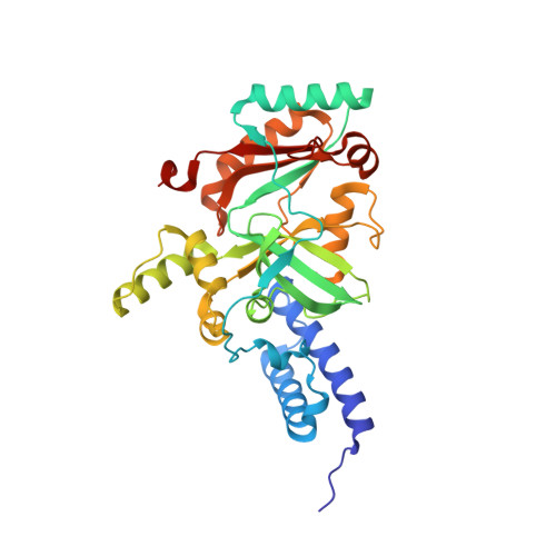
4LH6

Crystal structure of a LigA inhibitor


Experimental Data Snapshot

  • Method: X-RAY DIFFRACTION
  • Resolution: 1.65 Å
  • R-Value Free: 0.248 
  • R-Value Work: 0.219 
  • R-Value Observed: 0.221 

wwPDB Validation   3D Report Full Report


Ligand Structure Quality Assessment 


This is version 1.2 of the entry. See complete history


Literature

Identification through structure-based methods of a bacterial NAD(+)-dependent DNA ligase inhibitor that avoids known resistance mutations.

Murphy-Benenato, K.Wang, H.McGuire, H.M.Davis, H.E.Gao, N.Prince, D.B.Jahic, H.Stokes, S.S.Boriack-Sjodin, P.A.

(2014) Bioorg Med Chem Lett 24: 360-366

  • DOI: https://doi.org/10.1016/j.bmcl.2013.11.007
  • Primary Citation of Related Structures:  
    4LH6, 4LH7

  • PubMed Abstract: 

    In an attempt to identify novel inhibitors of NAD(+)-dependent DNA ligase (LigA) that are not affected by a known resistance mutation in the adenosine binding pocket, a detailed analysis of the binding sites of a variety of bacterial ligases was performed. This analysis revealed several similarities to the adenine binding region of kinases, which enabled a virtual screen of known kinase inhibitors. From this screen, a thienopyridine scaffold was identified that was shown to inhibit bacterial ligase. Further characterization through structure and enzymology revealed the compound was not affected by a previously disclosed resistance mutation in Streptococcus pneumoniae LigA, Leu75Phe. A subsequent medicinal chemistry program identified substitutions that resulted in an inhibitor with moderate activity across various Gram-positive bacterial LigA enzymes.


  • Organizational Affiliation

    Department of Chemistry, Infection Innovative Medicines, AstraZeneca R&D Boston, 35 Gatehouse Dr., Waltham, MA 02451, United States. Electronic address: kerry.benenato@astrazeneca.com.


Macromolecules
Find similar proteins by:  (by identity cutoff)  |  3D Structure
Entity ID: 1
MoleculeChains Sequence LengthOrganismDetailsImage
DNA ligase323Enterococcus faecalis V583Mutation(s): 0 
Gene Names: ligAEF_0722
EC: 6.5.1.2
UniProt
Find proteins for Q837V6 (Enterococcus faecalis (strain ATCC 700802 / V583))
Explore Q837V6 
Go to UniProtKB:  Q837V6
Entity Groups  
Sequence Clusters30% Identity50% Identity70% Identity90% Identity95% Identity100% Identity
UniProt GroupQ837V6
Sequence Annotations
Expand
  • Reference Sequence
Small Molecules
Binding Affinity Annotations 
IDSourceBinding Affinity
1X7 PDBBind:  4LH6 IC50: 7.70e+4 (nM) from 1 assay(s)
Experimental Data & Validation

Experimental Data

  • Method: X-RAY DIFFRACTION
  • Resolution: 1.65 Å
  • R-Value Free: 0.248 
  • R-Value Work: 0.219 
  • R-Value Observed: 0.221 
  • Space Group: C 2 2 21
Unit Cell:
Length ( Å )Angle ( ˚ )
a = 48.955α = 90
b = 104.377β = 90
c = 136.709γ = 90
Software Package:
Software NamePurpose
CrystalCleardata collection
AMoREphasing
REFMACrefinement
d*TREKdata reduction
d*TREKdata scaling

Structure Validation

View Full Validation Report


Ligand Structure Quality Assessment 


Entry History 

Deposition Data

Revision History  (Full details and data files)

  • Version 1.0: 2013-12-25
    Type: Initial release
  • Version 1.1: 2014-01-08
    Changes: Database references
  • Version 1.2: 2024-02-28
    Changes: Data collection, Database references, Derived calculations