4LH2

Structure of mouse 1-Pyrroline-5-Carboxylate Dehydrogenase (ALDH4A1) complexed with succinate


Experimental Data Snapshot

  • Method: X-RAY DIFFRACTION
  • Resolution: 1.67 Å
  • R-Value Free: 0.189 
  • R-Value Work: 0.164 
  • R-Value Observed: 0.165 

wwPDB Validation   3D Report Full Report


This is version 1.2 of the entry. See complete history


Literature

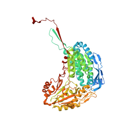
Structural basis of substrate selectivity of Delta (1)-pyrroline-5-carboxylate dehydrogenase (ALDH4A1): Semialdehyde chain length.

Pemberton, T.A.Tanner, J.J.

(2013) Arch Biochem Biophys 538: 34-40

  • DOI: https://doi.org/10.1016/j.abb.2013.07.024
  • Primary Citation of Related Structures:  
    4LGZ, 4LH0, 4LH1, 4LH2, 4LH3

  • PubMed Abstract: 

    The enzyme Δ(1)-pyrroline-5-carboxylate (P5C) dehydrogenase (aka P5CDH and ALDH4A1) is an aldehyde dehydrogenase that catalyzes the oxidation of γ-glutamate semialdehyde to l-glutamate. The crystal structures of mouse P5CDH complexed with glutarate, succinate, malonate, glyoxylate, and acetate are reported. The structures are used to build a structure-activity relationship that describes the semialdehyde carbon chain length and the position of the aldehyde group in relation to the cysteine nucleophile and oxyanion hole. Efficient 4- and 5-carbon substrates share the common feature of being long enough to span the distance between the anchor loop at the bottom of the active site and the oxyanion hole at the top of the active site. The inactive 2- and 3-carbon semialdehydes bind the anchor loop but are too short to reach the oxyanion hole. Inhibition of P5CDH by glyoxylate, malonate, succinate, glutarate, and l-glutamate is also examined. The Ki values are 0.27 mM for glyoxylate, 58 mM for succinate, 30 mM for glutarate, and 12 mM for l-glutamate. Curiously, malonate is not an inhibitor. The trends in Ki likely reflect a trade-off between the penalty for desolvating the carboxylates of the free inhibitor and the number of compensating hydrogen bonds formed in the enzyme-inhibitor complex.


  • Organizational Affiliation

    Department of Chemistry, University of Missouri-Columbia, Columbia, MO 65211, USA.


Macromolecules
Find similar proteins by:  (by identity cutoff)  |  3D Structure
Entity ID: 1
MoleculeChains Sequence LengthOrganismDetailsImage
Delta-1-pyrroline-5-carboxylate dehydrogenase, mitochondrial
A, B
563Mus musculusMutation(s): 0 
Gene Names: ADLH4a1Aldh4a1
EC: 1.5.1.12
UniProt
Find proteins for Q8CHT0 (Mus musculus)
Explore Q8CHT0 
Go to UniProtKB:  Q8CHT0
Entity Groups  
Sequence Clusters30% Identity50% Identity70% Identity90% Identity95% Identity100% Identity
UniProt GroupQ8CHT0
Sequence Annotations
Expand
  • Reference Sequence
Small Molecules
Binding Affinity Annotations 
IDSourceBinding Affinity
SIN Binding MOAD:  4LH2 Ki: 5.80e+7 (nM) from 1 assay(s)
PDBBind:  4LH2 Ki: 5.80e+7 (nM) from 1 assay(s)
Experimental Data & Validation

Experimental Data

  • Method: X-RAY DIFFRACTION
  • Resolution: 1.67 Å
  • R-Value Free: 0.189 
  • R-Value Work: 0.164 
  • R-Value Observed: 0.165 
  • Space Group: P 21 21 21
Unit Cell:
Length ( Å )Angle ( ˚ )
a = 84.884α = 90
b = 94.091β = 90
c = 132.339γ = 90
Software Package:
Software NamePurpose
SCALAdata scaling
PHENIXrefinement
PDB_EXTRACTdata extraction
CrystalCleardata collection
XDSdata reduction
PHENIXphasing

Structure Validation

View Full Validation Report


Entry History 

Deposition Data

Revision History  (Full details and data files)

  • Version 1.0: 2013-08-28
    Type: Initial release
  • Version 1.1: 2013-10-09
    Changes: Database references
  • Version 1.2: 2023-09-20
    Changes: Data collection, Database references, Derived calculations, Refinement description