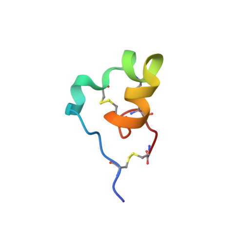
Native Chemical Ligation at Asx-Cys, Glx-Cys: Chemical Synthesis and High-Resolution X-ray Structure of ShK Toxin by Racemic Protein Crystallography.
Dang, B., Kubota, T., Mandal, K., Bezanilla, F., Kent, S.B.(2013) J Am Chem Soc 135: 11911-11919
- PubMed: 23919482 Search on PubMedSearch on PubMed Central
- DOI: https://doi.org/10.1021/ja4046795
- Primary Citation Related Structures:
4LFQ, 4LFS - PubMed Abstract:
We have re-examined the utility of native chemical ligation at -Gln/Glu-Cys- [Glx-Cys] and -Asn/Asp-Cys- [Asx-Cys] sites. Using the improved thioaryl catalyst 4-mercaptophenylacetic acid (MPAA), native chemical ligation could be performed at -Gln-Cys- and Asn-Cys- sites without side reactions. After optimization, ligation at a -Glu-Cys- site could also be used as a ligation site, with minimal levels of byproduct formation. However, -Asp-Cys- is not appropriate for use as a site for native chemical ligation because of formation of significant amounts of β-linked byproduct. The feasibility of native chemical ligation at -Gln-Cys- enabled a convergent total chemical synthesis of the enantiomeric forms of the ShK toxin protein molecule. The D-ShK protein molecule was ~50,000-fold less active in blocking the Kv1.3 channel than the L-ShK protein molecule. Racemic protein crystallography was used to obtain high-resolution X-ray diffraction data for ShK toxin. The structure was solved by direct methods and showed significant differences from the previously reported NMR structures in some regions of the ShK protein molecule.
- Department of Chemistry, Institute for Biophysical Dynamics, University of Chicago, Chicago, Illinois 60637, USA.
Organizational Affiliation: