4LEZ

Structure of mouse cGAS bound to an 18bp DNA and cGAS product


Experimental Data Snapshot

  • Method: X-RAY DIFFRACTION
  • Resolution: 2.36 Å
  • R-Value Free: 0.247 
  • R-Value Work: 0.192 
  • R-Value Observed: 0.195 

wwPDB Validation   3D Report Full Report


Ligand Structure Quality Assessment 


This is version 1.2 of the entry. See complete history


Literature

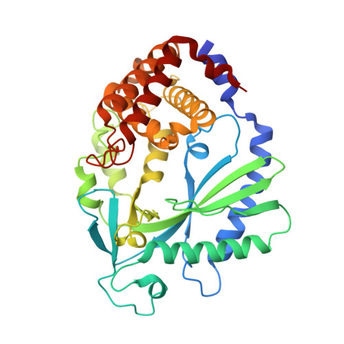
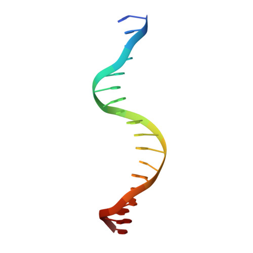
Cyclic GMP-AMP Synthase Is Activated by Double-Stranded DNA-Induced Oligomerization.

Li, X.Shu, C.Yi, G.Chaton, C.T.Shelton, C.L.Diao, J.Zuo, X.Kao, C.C.Herr, A.B.Li, P.

(2013) Immunity 39: 1019-1031

  • DOI: https://doi.org/10.1016/j.immuni.2013.10.019
  • Primary Citation of Related Structures:  
    4LEV, 4LEW, 4LEY, 4LEZ

  • PubMed Abstract: 
  • Cyclic GMP-AMP synthase (cGAS) is a cytosolic DNA sensor mediating innate antimicrobial immunity. It catalyzes the synthesis of a noncanonical cyclic dinucleotide, 2',5' cGAMP, that binds to STING and mediates the activation of TBK1 and IRF-3. Activated IRF-3 translocates to the nucleus and initiates the transcription of the IFN-β gene ...

    Cyclic GMP-AMP synthase (cGAS) is a cytosolic DNA sensor mediating innate antimicrobial immunity. It catalyzes the synthesis of a noncanonical cyclic dinucleotide, 2',5' cGAMP, that binds to STING and mediates the activation of TBK1 and IRF-3. Activated IRF-3 translocates to the nucleus and initiates the transcription of the IFN-β gene. The structure of mouse cGAS bound to an 18 bp dsDNA revealed that cGAS interacts with dsDNA through two binding sites, forming a 2:2 complex. Enzyme assays and IFN-β reporter assays of cGAS mutants demonstrated that interactions at both DNA binding sites are essential for cGAS activation. Mutagenesis and DNA binding studies showed that the two sites bind dsDNA cooperatively and that site B plays a critical role in DNA binding. The structure of mouse cGAS bound to dsDNA and 2',5' cGAMP provided insight into the catalytic mechanism of cGAS. These results demonstrated that cGAS is activated by dsDNA-induced oligomerization.


    Organizational Affiliation

    Department of Biochemistry and Biophysics, Texas A&M University, College Station, TX 77843-2128, USA. Electronic address: pingwei@tamu.edu.


Macromolecules

Find similar proteins by:  (by identity cutoff)  |  3D Structure
Entity ID: 1
MoleculeChainsSequence LengthOrganismDetailsImage
Cyclic GMP-AMP synthaseA,
B [auth C]
366Mus musculusMutation(s): 0 
Gene Names: Mb21d1Cgas
EC: 2.7.7 (PDB Primary Data), 2.7.7.86 (UniProt)
UniProt & NIH Common Fund Data Resources
Find proteins for Q8C6L5 (Mus musculus)
Explore Q8C6L5 
Go to UniProtKB:  Q8C6L5
IMPC:  MGI:2442261
Entity Groups  
Sequence Clusters30% Identity50% Identity70% Identity90% Identity95% Identity100% Identity
UniProt GroupQ8C6L5
Protein Feature View
Expand
  • Reference Sequence

Find similar nucleic acids by:  Sequence   |   3D Structure  

Entity ID: 2
MoleculeChainsLengthOrganismImage
18bp dsDNAC [auth E],
D [auth F],
E [auth I],
F [auth J]
18N/A
Protein Feature View
Expand
  • Reference Sequence
Small Molecules
Ligands 2 Unique
IDChainsName / Formula / InChI Key2D Diagram3D Interactions
1SY
Query on 1SY

Download Ideal Coordinates CCD File 
H [auth A],
J [auth C]
cGAMP
C20 H24 N10 O13 P2
XRILCFTWUCUKJR-INFSMZHSSA-N
 Ligand Interaction
ZN
Query on ZN

Download Ideal Coordinates CCD File 
G [auth A],
I [auth C]
ZINC ION
Zn
PTFCDOFLOPIGGS-UHFFFAOYSA-N
 Ligand Interaction
Experimental Data & Validation

Experimental Data

  • Method: X-RAY DIFFRACTION
  • Resolution: 2.36 Å
  • R-Value Free: 0.247 
  • R-Value Work: 0.192 
  • R-Value Observed: 0.195 
  • Space Group: P 21 21 21
Unit Cell:
Length ( Å )Angle ( ˚ )
a = 79.209α = 90
b = 99.031β = 90
c = 142.631γ = 90
Software Package:
Software NamePurpose
CrystalCleardata collection
PHASERphasing
PHENIXrefinement
HKL-2000data reduction
HKL-2000data scaling

Structure Validation

View Full Validation Report


Ligand Structure Quality Assessment 


Entry History 

Deposition Data

  • Released Date: 2013-12-25 
  • Deposition Author(s): Li, P.

Revision History  (Full details and data files)

  • Version 1.0: 2013-12-25
    Type: Initial release
  • Version 1.1: 2014-01-08
    Changes: Database references
  • Version 1.2: 2015-08-05
    Changes: Non-polymer description