The crystal structures of the tri-functional Chloroflexus aurantiacus and bi-functional Rhodobacter sphaeroides malyl-CoA lyases and comparison with CitE-like superfamily enzymes and malate synthases.
Zarzycki, J., Kerfeld, C.A.(2013) BMC Struct Biol 13: 28-28
- PubMed: 24206647 Search on PubMedSearch on PubMed Central
- DOI: https://doi.org/10.1186/1472-6807-13-28
- Primary Citation Related Structures:
4L7Z, 4L80, 4L9Y, 4L9Z - PubMed Abstract:
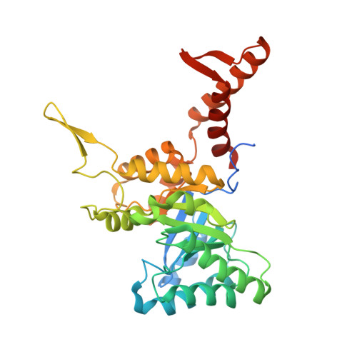
Malyl-CoA lyase (MCL) is a promiscuous carbon-carbon bond lyase that catalyzes the reversible cleavage of structurally related Coenzyme A (CoA) thioesters. This enzyme plays a crucial, multifunctional role in the 3-hydroxypropionate bi-cycle for autotrophic CO2 fixation in Chloroflexus aurantiacus. A second, phylogenetically distinct MCL from Rhodobacter sphaeroides is involved in the ethylmalonyl-CoA pathway for acetate assimilation. Both MCLs belong to the large superfamily of CitE-like enzymes, which includes the name-giving β-subunit of citrate lyase (CitE), malyl-CoA thioesterases and other enzymes of unknown physiological function. The CitE-like enzyme superfamily also bears sequence and structural resemblance to the malate synthases. All of these different enzymes share highly conserved catalytic residues, although they catalyze distinctly different reactions: C-C bond formation and cleavage, thioester hydrolysis, or both (the malate synthases). Here we report the first crystal structures of MCLs from two different phylogenetic subgroups in apo- and substrate-bound forms. Both the C. aurantiacus and the R. sphaeroides MCL contain elaborations on the canonical β8/α8 TIM barrel fold and form hexameric assemblies. Upon ligand binding, changes in the C-terminal domains of the MCLs result in closing of the active site, with the C-terminal domain of one monomer forming a lid over and contributing side chains to the active site of the adjacent monomer. The distinctive features of the two MCL subgroups were compared to known structures of other CitE-like superfamily enzymes and to malate synthases, providing insight into the structural subtleties that underlie the functional versatility of these enzymes. Although the C. aurantiacus and the R. sphaeroides MCLs have divergent primary structures (~37% identical), their tertiary and quaternary structures are very similar. It can be assumed that the C-C bond formation catalyzed by the MCLs occurs as proposed for malate synthases. However, a comparison of the two MCL structures with known malate synthases raised the question why the MCLs are not also able to hydrolyze CoA thioester bonds. Our results suggest the previously proposed reaction mechanism for malate synthases may be incomplete or not entirely correct. Further studies involving site-directed mutagenesis based on these structures may be required to solve this puzzling question.
- Department of Biochemistry and Molecular Biology, Plant Research Laboratories, Michigan State University, Plant Biology Building, 612 Wilson Road, East Lansing, MI 48824, USA. ckerfeld@lbl.gov.
Organizational Affiliation: