Structural and functional characterization of a novel alpha / beta hydrolase from cariogenic pathogen Streptococcus mutans.
Wang, Z., Li, L., Su, X.D.(2014) Proteins 82: 695-700
- PubMed: 24115105 Search on PubMed
- DOI: https://doi.org/10.1002/prot.24418
- Primary Citation Related Structures:
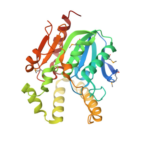
4L9A - PubMed Abstract:
The protein Smu.1393c from Streptococcus mutans is annotated as a putative α/β hydrolase, but it has low sequence identity to the structure-known α/β hydrolases. Here we present the crystal structure of Smu.1393c at 2.0 Å resolution. Smu.1393c has a fully open alkaline substrate pocket, whose conformation is unique among other similar hydrolase structures. Three residues, Ser101, His251, and Glu125, were identified as the active center of Smu.1393c. By screening a series of artificial hydrolase substrates, we demonstrated Smu.1393c had low carboxylesterase activity towards short-chain carboxyl esters, which provided a clue for exploring the in vivo function of Smu.1393c.
- State Key Laboratory of Protein and Plant Gene Research, and Biodynamic Optical Imaging Center (BIOPIC), School of Life Sciences, Peking University, Beijing, 100871, China.
Organizational Affiliation: