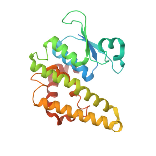
4L8E

Crystal structure of a glutathione transferase family member from xenorhabdus nematophila, target efi-507418, with two gsh per subunit


Experimental Data Snapshot

  • Method: X-RAY DIFFRACTION
  • Resolution: 1.70 Å
  • R-Value Free: 0.176 
  • R-Value Work: 0.151 
  • R-Value Observed: 0.152 

wwPDB Validation   3D Report Full Report


Ligand Structure Quality Assessment 


This is version 1.1 of the entry. See complete history


Literature

Crystal structure of a glutathione transferase family member from xenorhabdus nematophila, target efi-507418, with two gsh per subunit

Vetting, M.W.Toro, R.Bhosle, R.Al Obaidi, N.F.Morisco, L.L.Wasserman, S.R.Sojitra, S.Stead, M.Washington, E.Scott Glenn, A.Chowdhury, S.Evans, B.Hammonds, J.Hillerich, B.Love, J.Seidel, R.D.Imker, H.J.Gerlt, J.A.Armstrong, R.N.Almo, S.C.Enzyme Function Initiative (EFI)

To be published.

Macromolecules
Find similar proteins by:  (by identity cutoff)  |  3D Structure
Entity ID: 1
MoleculeChainsSequence LengthOrganismDetailsImage
Glutathione S-transferase enzyme with thioredoxin-like domain235Xenorhabdus nematophila ATCC 19061Mutation(s): 0 
Gene Names: yfcGXNC1_3157
UniProt
Find proteins for D3VKV1 (Xenorhabdus nematophila (strain ATCC 19061 / DSM 3370 / CCUG 14189 / LMG 1036 / NCIMB 9965 / AN6))
Explore D3VKV1 
Go to UniProtKB:  D3VKV1
Entity Groups  
Sequence Clusters30% Identity50% Identity70% Identity90% Identity95% Identity100% Identity
UniProt GroupD3VKV1
Protein Feature View
Expand
  • Reference Sequence
Small Molecules
Ligands 3 Unique
IDChainsName / Formula / InChI Key2D Diagram3D Interactions
GSH
Query on GSH

Download Ideal Coordinates CCD File 
B [auth A],
C [auth A],
D [auth A]
GLUTATHIONE
C10 H17 N3 O6 S
RWSXRVCMGQZWBV-WDSKDSINSA-N
 Ligand Interaction
MES
Query on MES

Download Ideal Coordinates CCD File 
F [auth A]2-(N-MORPHOLINO)-ETHANESULFONIC ACID
C6 H13 N O4 S
SXGZJKUKBWWHRA-UHFFFAOYSA-N
 Ligand Interaction
CL
Query on CL

Download Ideal Coordinates CCD File 
E [auth A]CHLORIDE ION
Cl
VEXZGXHMUGYJMC-UHFFFAOYSA-M
 Ligand Interaction
Experimental Data & Validation

Experimental Data

  • Method: X-RAY DIFFRACTION
  • Resolution: 1.70 Å
  • R-Value Free: 0.176 
  • R-Value Work: 0.151 
  • R-Value Observed: 0.152 
  • Space Group: P 65 2 2
Unit Cell:
Length ( Å )Angle ( ˚ )
a = 65.331α = 90
b = 65.331β = 90
c = 213.919γ = 120
Software Package:
Software NamePurpose
SCALAdata scaling
PHENIXrefinement
PDB_EXTRACTdata extraction
MOSFLMdata reduction
AMoREphasing

Structure Validation

View Full Validation Report


Ligand Structure Quality Assessment 


Entry History 

Revision History  (Full details and data files)

  • Version 1.0: 2013-06-26
    Type: Initial release
  • Version 1.1: 2023-09-20
    Changes: Data collection, Database references, Derived calculations, Refinement description