4L5O

Crystal structure of 26 kDa GST D26H mutant of Clonorchis sinensis


Experimental Data Snapshot

  • Method: X-RAY DIFFRACTION
  • Resolution: 2.09 Å
  • R-Value Free: 0.248 
  • R-Value Work: 0.202 
  • R-Value Observed: 0.204 

wwPDB Validation   3D Report Full Report


Ligand Structure Quality Assessment 


This is version 1.2 of the entry. See complete history


Literature

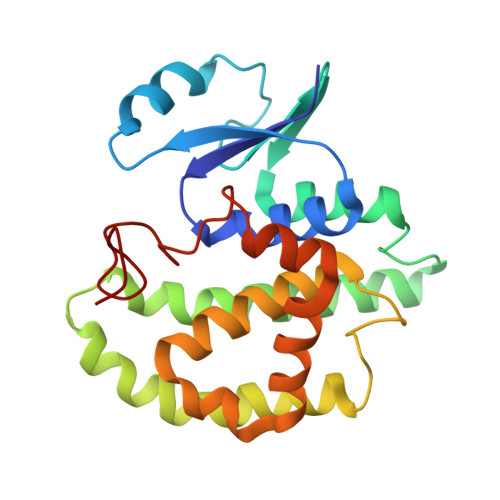
Crystal structures of 26kDa Clonorchis sinensis glutathione S-transferase reveal zinc binding and putative metal binding.

Han, Y.H.Hong, S.J.Cheong, H.K.Chung, Y.J.

(2013) Biochem Biophys Res Commun 438: 457-461

  • DOI: https://doi.org/10.1016/j.bbrc.2013.07.102
  • Primary Citation of Related Structures:  
    4L5L, 4L5O

  • PubMed Abstract: 
  • The crystal structures of CsGST in two different space groups revealed that Asp26 and His79 coordinate a zinc ion. In one space group, His46 of an adjacent molecule participates in the coordination within 2.0Å. In the other space group, Asp26, His79 and a water molecule coordinate a zinc ion ...

    The crystal structures of CsGST in two different space groups revealed that Asp26 and His79 coordinate a zinc ion. In one space group, His46 of an adjacent molecule participates in the coordination within 2.0Å. In the other space group, Asp26, His79 and a water molecule coordinate a zinc ion. The CsGST-D26H structure showed that four histidine residues - His26 and His79 from one molecule and the same residues from a symmetry-related neighboring molecule - coordinate a zinc ion. The coordinated zinc ions are located between two molecules and mediate molecular contacts within the crystal.


    Organizational Affiliation

    Division of Magnetic Resonance Research, Korea Basic Science Institute (KBSI), Ochang, Chungbuk 363-883, Republic of Korea.


Macromolecules
Find similar proteins by:  (by identity cutoff)  |  3D Structure
Entity ID: 1
MoleculeChainsSequence LengthOrganismDetailsImage
Putative glutathione transferase
A, B, C
218Clonorchis sinensisMutation(s): 1 
Gene Names: CSKR_11935s
EC: 2.5.1.18
Entity Groups  
Sequence Clusters30% Identity50% Identity70% Identity90% Identity95% Identity100% Identity
Protein Feature View
Expand
  • Reference Sequence
Experimental Data & Validation

Experimental Data

  • Method: X-RAY DIFFRACTION
  • Resolution: 2.09 Å
  • R-Value Free: 0.248 
  • R-Value Work: 0.202 
  • R-Value Observed: 0.204 
  • Space Group: P 31 2 1
Unit Cell:
Length ( Å )Angle ( ˚ )
a = 98.37α = 90
b = 98.37β = 90
c = 178.501γ = 120
Software Package:
Software NamePurpose
SCALAdata scaling
MOLREPphasing
REFMACrefinement
PDB_EXTRACTdata extraction
HKL-2000data collection
DENZOdata reduction
SCALEPACKdata scaling

Structure Validation

View Full Validation Report


Ligand Structure Quality Assessment 


Entry History 

Deposition Data

Revision History  (Full details and data files)

  • Version 1.0: 2013-09-11
    Type: Initial release
  • Version 1.1: 2015-06-17
    Changes: Database references
  • Version 1.2: 2017-11-15
    Changes: Refinement description