Pyridone-conjugated monobactam antibiotics with gram-negative activity.
Brown, M.F., Mitton-Fry, M.J., Arcari, J.T., Barham, R., Casavant, J., Gerstenberger, B.S., Han, S., Hardink, J.R., Harris, T.M., Hoang, T., Huband, M.D., Lall, M.S., Lemmon, M.M., Li, C., Lin, J., McCurdy, S.P., McElroy, E., McPherson, C., Marr, E.S., Mueller, J.P., Mullins, L., Nikitenko, A.A., Noe, M.C., Penzien, J., Plummer, M.S., Schuff, B.P., Shanmugasundaram, V., Starr, J.T., Sun, J., Tomaras, A., Young, J.A., Zaniewski, R.P.(2013) J Med Chem 56: 5541-5552
- PubMed: 23755848 Search on PubMed
- DOI: https://doi.org/10.1021/jm400560z
- Primary Citation Related Structures:
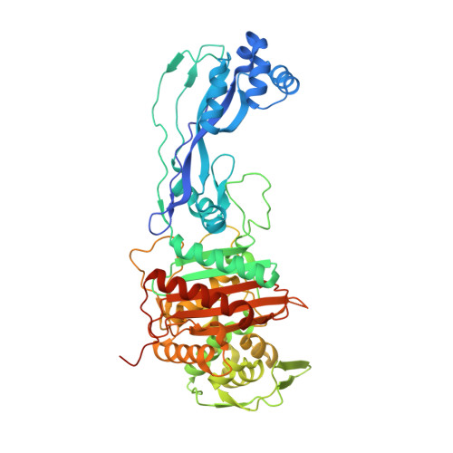
4L0L - PubMed Abstract:
Herein we describe the structure-aided design and synthesis of a series of pyridone-conjugated monobactam analogues with in vitro antibacterial activity against clinically relevant Gram-negative species including Pseudomonas aeruginosa , Klebsiella pneumoniae , and Escherichia coli . Rat pharmacokinetic studies with compound 17 demonstrate low clearance and low plasma protein binding. In addition, evidence is provided for a number of analogues suggesting that the siderophore receptors PiuA and PirA play a role in drug uptake in P. aeruginosa strain PAO1.
- Worldwide Medicinal Chemistry, ‡Computational Chemistry, §Antibacterials Research Unit, ∥Pharmacokinetics, Dynamics & Metabolism, ⊥Structural Biology, Pfizer Global Research and Development , Eastern Point Road, Groton, Connecticut 06340, United States.
Organizational Affiliation: