4KTQ | pdb_00004ktq

BINARY COMPLEX OF THE LARGE FRAGMENT OF DNA POLYMERASE I FROM T. AQUATICUS BOUND TO A PRIMER/TEMPLATE DNA


Experimental Data Snapshot

  • Method: X-RAY DIFFRACTION
  • Resolution: 2.50 Å
  • R-Value Free: 
    0.298 (Depositor) 
  • R-Value Work: 
    0.227 (Depositor) 
  • R-Value Observed: 
    0.227 (Depositor) 

wwPDB Validation 3D Report Full Report

Validation slider image for 4KTQ

This is version 1.3 of the entry. See complete history

Literature

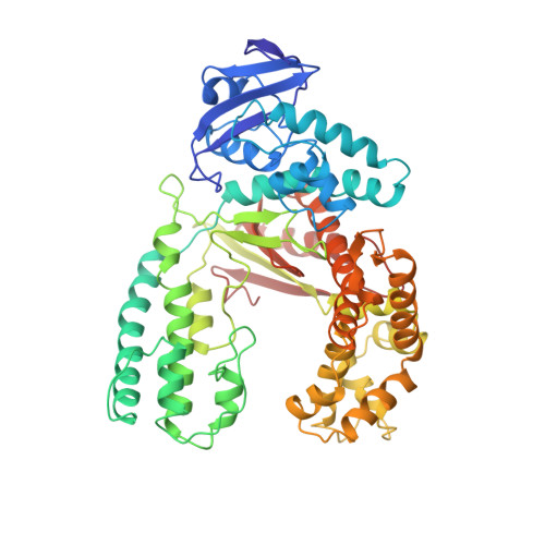
Crystal structures of open and closed forms of binary and ternary complexes of the large fragment of Thermus aquaticus DNA polymerase I: structural basis for nucleotide incorporation.

Li, Y.Korolev, S.Waksman, G.

(1998) EMBO J 17: 7514-7525

  • DOI: https://doi.org/10.1093/emboj/17.24.7514
  • Primary Citation Related Structures: 
    2KTQ, 3KTQ, 4KTQ

  • PubMed Abstract: 

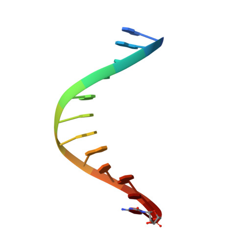
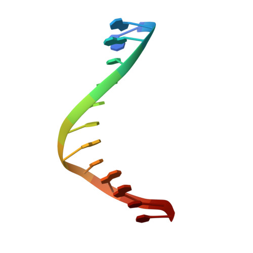
    The crystal structures of two ternary complexes of the large fragment of Thermus aquaticus DNA polymerase I (Klentaq1) with a primer/template DNA and dideoxycytidine triphosphate, and that of a binary complex of the same enzyme with a primer/template DNA, were determined to a resolution of 2.3, 2.3 and 2.5 A, respectively. One ternary complex structure differs markedly from the two other structures by a large reorientation of the tip of the fingers domain. This structure, designated 'closed', represents the ternary polymerase complex caught in the act of incorporating a nucleotide. In the two other structures, the tip of the fingers domain is rotated outward by 46 degrees ('open') in an orientation similar to that of the apo form of Klentaq1. These structures provide the first direct evidence in DNA polymerase I enzymes of a large conformational change responsible for assembling an active ternary complex.


  • Organizational Affiliation
    • Department of Biochemistry and Molecular Biophysics, Washington University School of Medicine, Campus Box 8231, 660 South Euclid Avenue, St Louis, MO 63110, USA.

Macromolecule Content 

  • Total Structure Weight: 68.51 kDa 
  • Atom Count: 4,769 
  • Modeled Residue Count: 564 
  • Deposited Residue Count: 564 
  • Unique protein chains: 1
  • Unique nucleic acid chains: 2

Macromolecules


Find similar proteins by:|  3D Structure
Entity ID: 3
MoleculeChains  Sequence LengthOrganismDetailsImage
PROTEIN (LARGE FRAGMENT OF DNA POLYMERASE I)C [auth A]539Thermus aquaticusMutation(s): 0 
Gene Names: TAQ
EC: 2.7.7.7
UniProt
Find proteins for P19821 (Thermus aquaticus)
Explore P19821 
Go to UniProtKB:  P19821
Entity Groups
Sequence Clusters30% Identity50% Identity70% Identity90% Identity95% Identity100% Identity
UniProt GroupP19821
Sequence Annotations
Expand
Reference Sequence
Find similar nucleic acids by:  Sequence
Entity ID: 1
MoleculeChains LengthOrganismImage
DNA (5'-D(*GP*AP*CP*CP*AP*CP*GP*GP*CP*GP*CP*(DOC))-3')A [auth B]12N/A
Sequence Annotations
Expand
Reference Sequence
Find similar nucleic acids by:  Sequence
Entity ID: 2
MoleculeChains LengthOrganismImage
DNA (5'-D(*GP*GP*GP*CP*GP*CP*CP*GP*TP*GP*GP*TP*C)-3')B [auth C]13N/A
Sequence Annotations
Expand
Reference Sequence

Experimental Data & Validation

Experimental Data

  • Method: X-RAY DIFFRACTION
  • Resolution: 2.50 Å
  • R-Value Free:  0.298 (Depositor) 
  • R-Value Work:  0.227 (Depositor) 
  • R-Value Observed: 0.227 (Depositor) 
Space Group: P 31 2 1
Unit Cell:
Length ( Å )Angle ( ˚ )
a = 110.842α = 90
b = 110.842β = 90
c = 90.843γ = 120
Software Package:
Software NamePurpose
X-PLORrefinement

Structure Validation

View Full Validation Report


Entry History 

Deposition Data

Revision History  (Full details and data files)

  • Version 1.0: 1999-01-13
    Type: Initial release
  • Version 1.1: 2008-04-27
    Changes: Version format compliance
  • Version 1.2: 2011-07-13
    Changes: Version format compliance
  • Version 1.3: 2024-02-28
    Changes: Data collection, Database references, Derived calculations