4KP6 | pdb_00004kp6

Crystal structure of human phosphodiesterase 4B (PDE4B) in complex with a [1,3,5]triazine derivative


Experimental Data Snapshot

  • Method: X-RAY DIFFRACTION
  • Resolution: 1.50 Å
  • R-Value Free: 
    0.198 (Depositor), 0.205 (DCC) 
  • R-Value Work: 
    0.168 (Depositor), 0.175 (DCC) 
  • R-Value Observed: 
    0.169 (Depositor) 

wwPDB Validation 3D Report Full Report

Validation slider image for 4KP6

Ligand Structure Quality Assessment 


This is version 1.3 of the entry. See complete history

Literature

Discovery of triazines as potent, selective and orally active PDE4 inhibitors.

Gewald, R.Grunwald, C.Egerland, U.

(2013) Bioorg Med Chem Lett 23: 4308-4314

  • DOI: https://doi.org/10.1016/j.bmcl.2013.05.099
  • Primary Citation Related Structures: 
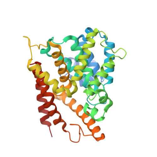
    4KP6

  • PubMed Abstract: 

    Expanding on HTS hit 4 afforded a series of [1,3,5]triazine derivatives as novel PDE4 inhibitors. The SAR development and optimization process with the emphasis on ligand efficiency and physicochemical properties led to the discovery of compound 44 as a potent, selective and orally active PDE4 inhibitor.


  • Organizational Affiliation
    • BioCrea GmbH, Meissner Strasse 191, 01445 Radebeul, Germany. rainer.gewald@telecolumbus.net

Macromolecule Content 

  • Total Structure Weight: 40.59 kDa 
  • Atom Count: 2,933 
  • Modeled Residue Count: 324 
  • Deposited Residue Count: 340 
  • Unique protein chains: 1

Macromolecules

Find similar proteins by:|  3D Structure
Entity ID: 1
MoleculeChains  Sequence LengthOrganismDetailsImage
cAMP-specific 3',5'-cyclic phosphodiesterase 4B340Homo sapiensMutation(s): 0 
Gene Names: DPDE4PDE4B
EC: 3.1.4.17 (PDB Primary Data), 3.1.4.53 (UniProt)
UniProt & NIH Common Fund Data Resources
Find proteins for Q07343 (Homo sapiens)
Explore Q07343 
Go to UniProtKB:  Q07343
PHAROS:  Q07343
GTEx:  ENSG00000184588 
Entity Groups
Sequence Clusters30% Identity50% Identity70% Identity90% Identity95% Identity100% Identity
UniProt GroupQ07343
Sequence Annotations
Expand
Reference Sequence

Small Molecules

Ligands 4 Unique
IDChains Name / Formula / InChI Key2D Diagram3D Interactions
1S1

Query on 1S1


Download:Ideal Coordinates CCD File
B [auth A]2-ethyl-2-{[4-(methylamino)-6-(1H-1,2,4-triazol-1-yl)-1,3,5-triazin-2-yl]amino}butanenitrile
C12 H17 N9
AQTLNSKLZWRJEV-UHFFFAOYSA-N
ZN

Query on ZN


Download:Ideal Coordinates CCD File
D [auth A]ZINC ION
Zn
PTFCDOFLOPIGGS-UHFFFAOYSA-N
EDO

Query on EDO


Download:Ideal Coordinates CCD File
E [auth A]
F [auth A]
G [auth A]
H [auth A]
I [auth A]
E [auth A],
F [auth A],
G [auth A],
H [auth A],
I [auth A],
J [auth A],
K [auth A],
L [auth A],
M [auth A],
N [auth A],
O [auth A],
P [auth A],
Q [auth A],
R [auth A],
S [auth A],
T [auth A],
U [auth A],
V [auth A],
W [auth A]
1,2-ETHANEDIOL
C2 H6 O2
LYCAIKOWRPUZTN-UHFFFAOYSA-N
MG

Query on MG


Download:Ideal Coordinates CCD File
C [auth A]MAGNESIUM ION
Mg
JLVVSXFLKOJNIY-UHFFFAOYSA-N

Experimental Data & Validation

Experimental Data

  • Method: X-RAY DIFFRACTION
  • Resolution: 1.50 Å
  • R-Value Free:  0.198 (Depositor), 0.205 (DCC) 
  • R-Value Work:  0.168 (Depositor), 0.175 (DCC) 
  • R-Value Observed: 0.169 (Depositor) 
Space Group: P 43 21 2
Unit Cell:
Length ( Å )Angle ( ˚ )
a = 53.332α = 90
b = 53.332β = 90
c = 229.048γ = 90
Software Package:
Software NamePurpose
MAR345data collection
MOLREPphasing
REFMACrefinement
MOSFLMdata reduction
SCALAdata scaling

Structure Validation

View Full Validation Report


Ligand Structure Quality Assessment 


Entry History 

Deposition Data

Revision History  (Full details and data files)

  • Version 1.0: 2013-07-10
    Type: Initial release
  • Version 1.1: 2013-07-17
    Changes: Database references
  • Version 1.2: 2017-11-15
    Changes: Advisory, Refinement description
  • Version 1.3: 2024-02-28
    Changes: Advisory, Data collection, Database references, Derived calculations