4KML

Probing the N-terminal beta-sheet conversion in the crystal structure of the full-length human prion protein bound to a Nanobody


Experimental Data Snapshot

  • Method: X-RAY DIFFRACTION
  • Resolution: 1.50 Å
  • R-Value Free: 0.192 
  • R-Value Work: 0.175 
  • R-Value Observed: 0.176 

wwPDB Validation   3D Report Full Report


This is version 1.2 of the entry. See complete history


Literature

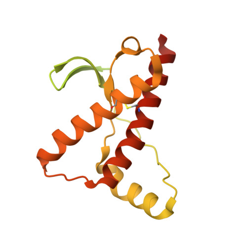
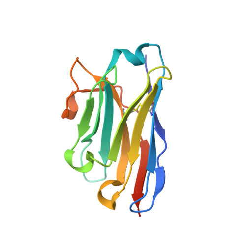
Probing the N-Terminal beta-Sheet Conversion in the Crystal Structure of the Human Prion Protein Bound to a Nanobody.

Abskharon, R.N.Giachin, G.Wohlkonig, A.Soror, S.H.Pardon, E.Legname, G.Steyaert, J.

(2014) J Am Chem Soc 136: 937-944

  • DOI: https://doi.org/10.1021/ja407527p
  • Primary Citation of Related Structures:  
    4KML, 4N9O

  • PubMed Abstract: 

    Prions are fatal neurodegenerative transmissible agents causing several incurable illnesses in humans and animals. Prion diseases are caused by the structural conversion of the cellular prion protein, PrP(C), into its misfolded oligomeric form, known as prion or PrP(Sc). The canonical human PrP(C) (HuPrP) fold features an unstructured N-terminal part (residues 23-124) and a well-defined C-terminal globular domain (residues 125-231). Compelling evidence indicates that an evolutionary N-terminal conserved motif AGAAAAGA (residues 113-120) plays an important role in the conversion to PrP(Sc). The intrinsic flexibility of the N-terminal has hampered efforts to obtain detailed atomic information on the structural features of this palindromic region. In this study, we crystallized the full-length HuPrP in complex with a nanobody (Nb484) that inhibits prion propagation. In the complex, the prion protein is unstructured from residue 23 to 116. The palindromic motif adopts a stable and fully extended configuration to form a three-stranded antiparallel β-sheet with the β1 and β2 strands, demonstrating that the full-length HuPrP(C) can adopt a more elaborate β0-β1-α1-β2-α2-α3 structural organization than the canonical β1-α1-β2-α2-α3 prion-like fold. From this structure, it appears that the palindromic motif mediates β-enrichment in the PrP(C) monomer as one of the early events in the conversion of PrP(C) into PrP(Sc).


  • Organizational Affiliation

    Structural Biology Brussels, Vrije Universiteit Brussel , Pleinlaan 2, 1050 Brussels, Belgium.


Macromolecules
Find similar proteins by:  (by identity cutoff)  |  3D Structure
Entity ID: 1
MoleculeChains Sequence LengthOrganismDetailsImage
Major prion protein241Homo sapiensMutation(s): 0 
Gene Names: PRIPPRNPPRP
UniProt & NIH Common Fund Data Resources
Find proteins for P04156 (Homo sapiens)
Explore P04156 
Go to UniProtKB:  P04156
PHAROS:  P04156
GTEx:  ENSG00000171867 
Entity Groups  
Sequence Clusters30% Identity50% Identity70% Identity90% Identity95% Identity100% Identity
UniProt GroupP04156
Sequence Annotations
Expand
  • Reference Sequence
Find similar proteins by:  (by identity cutoff)  |  3D Structure
Entity ID: 2
MoleculeChains Sequence LengthOrganismDetailsImage
Nanobody130Lama glamaMutation(s): 0 
Entity Groups  
Sequence Clusters30% Identity50% Identity70% Identity90% Identity95% Identity100% Identity
Sequence Annotations
Expand
  • Reference Sequence
Experimental Data & Validation

Experimental Data

  • Method: X-RAY DIFFRACTION
  • Resolution: 1.50 Å
  • R-Value Free: 0.192 
  • R-Value Work: 0.175 
  • R-Value Observed: 0.176 
  • Space Group: C 1 2 1
Unit Cell:
Length ( Å )Angle ( ˚ )
a = 131.442α = 90
b = 45.921β = 96.09
c = 44.964γ = 90
Software Package:
Software NamePurpose
HKL-3000data collection
PHASERphasing
REFMACrefinement
HKL-3000data reduction
SCALAdata scaling

Structure Validation

View Full Validation Report


Entry History 

Deposition Data

Revision History  (Full details and data files)

  • Version 1.0: 2014-02-19
    Type: Initial release
  • Version 1.1: 2018-01-24
    Changes: Structure summary
  • Version 1.2: 2023-09-20
    Changes: Data collection, Database references, Refinement description