4KLQ

Observing a DNA polymerase choose right from wrong.


Experimental Data Snapshot

  • Method: X-RAY DIFFRACTION
  • Resolution: 2.00 Å
  • R-Value Free: 0.267 
  • R-Value Work: 0.214 
  • R-Value Observed: 0.218 

wwPDB Validation   3D Report Full Report


Ligand Structure Quality Assessment 


This is version 1.2 of the entry. See complete history


Literature

Observing a DNA polymerase choose right from wrong.

Freudenthal, B.D.Beard, W.A.Shock, D.D.Wilson, S.H.

(2013) Cell 154: 157-168

  • DOI: https://doi.org/10.1016/j.cell.2013.05.048
  • Primary Citation of Related Structures:  
    4KLD, 4KLE, 4KLF, 4KLG, 4KLH, 4KLI, 4KLJ, 4KLL, 4KLM, 4KLO, 4KLQ, 4KLS, 4KLT, 4KLU, 4LVS

  • PubMed Abstract: 

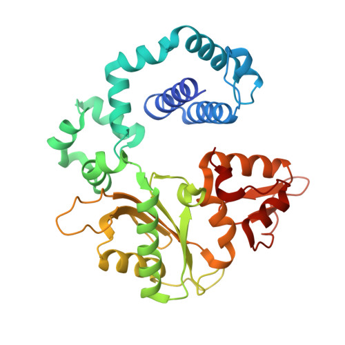
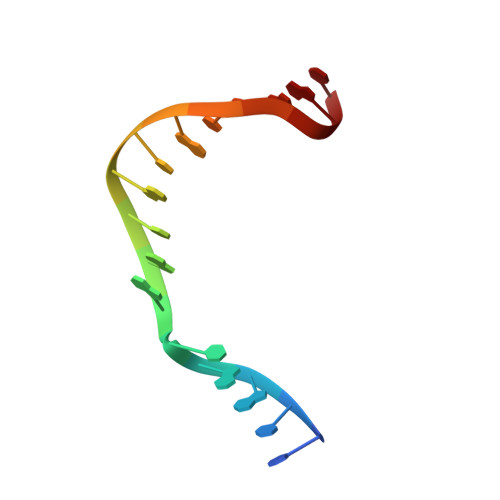
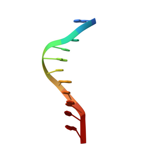
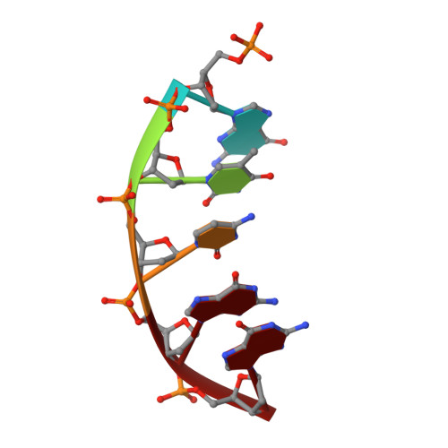
    DNA polymerase (pol) β is a model polymerase involved in gap-filling DNA synthesis utilizing two metals to facilitate nucleotidyl transfer. Previous structural studies have trapped catalytic intermediates by utilizing substrate analogs (dideoxy-terminated primer or nonhydrolysable incoming nucleotide). To identify additional intermediates during catalysis, we now employ natural substrates (correct and incorrect nucleotides) and follow product formation in real time with 15 different crystal structures. We are able to observe molecular adjustments at the active site that hasten correct nucleotide insertion and deter incorrect insertion not appreciated previously. A third metal binding site is transiently formed during correct, but not incorrect, nucleotide insertion. Additionally, long incubations indicate that pyrophosphate more easily dissociates after incorrect, compared to correct, nucleotide insertion. This appears to be coupled to subdomain repositioning that is required for catalytic activation/deactivation. The structures provide insights into a fundamental chemical reaction that impacts polymerase fidelity and genome stability.


  • Organizational Affiliation

    Laboratory of Structural Biology, National Institute of Environmental Health Sciences, National Institutes of Health, P.O. Box 12233, Research Triangle Park, NC 27709-2233, USA.


Macromolecules

Find similar proteins by:  (by identity cutoff)  |  3D Structure
Entity ID: 1
MoleculeChains Sequence LengthOrganismDetailsImage
DNA polymerase beta335Homo sapiensMutation(s): 0 
Gene Names: Polb
EC: 2.7.7.7 (PDB Primary Data), 4.2.99 (PDB Primary Data)
UniProt & NIH Common Fund Data Resources
Find proteins for P06746 (Homo sapiens)
Explore P06746 
Go to UniProtKB:  P06746
PHAROS:  P06746
GTEx:  ENSG00000070501 
Entity Groups  
Sequence Clusters30% Identity50% Identity70% Identity90% Identity95% Identity100% Identity
UniProt GroupP06746
Sequence Annotations
Expand
  • Reference Sequence

Find similar nucleic acids by:  Sequence   |   3D Structure  

Entity ID: 2
MoleculeChains LengthOrganismImage
5'-D(*CP*CP*GP*AP*CP*GP*GP*CP*GP*CP*AP*TP*CP*AP*GP*C)-3'B [auth T]16N/A
Sequence Annotations
Expand
  • Reference Sequence

Find similar nucleic acids by:  Sequence   |   3D Structure  

Entity ID: 3
MoleculeChains LengthOrganismImage
5'-D(*GP*CP*TP*GP*AP*TP*GP*CP*GP*CP*A)-3'C [auth P]11N/A
Sequence Annotations
Expand
  • Reference Sequence

Find similar nucleic acids by:  Sequence   |   3D Structure  

Entity ID: 4
MoleculeChains LengthOrganismImage
5'-D(P*GP*TP*CP*GP*G)-3'5N/A
Sequence Annotations
Expand
  • Reference Sequence
Small Molecules
Ligands 4 Unique
IDChains Name / Formula / InChI Key2D Diagram3D Interactions
DTP
Query on DTP

Download Ideal Coordinates CCD File 
E [auth A]2'-DEOXYADENOSINE 5'-TRIPHOSPHATE
C10 H16 N5 O12 P3
SUYVUBYJARFZHO-RRKCRQDMSA-N
PPV
Query on PPV

Download Ideal Coordinates CCD File 
F [auth A]PYROPHOSPHATE
H4 O7 P2
XPPKVPWEQAFLFU-UHFFFAOYSA-N
MN
Query on MN

Download Ideal Coordinates CCD File 
G [auth A],
H [auth A],
I [auth A]
MANGANESE (II) ION
Mn
WAEMQWOKJMHJLA-UHFFFAOYSA-N
NA
Query on NA

Download Ideal Coordinates CCD File 
J [auth A],
K [auth A],
L [auth A]
SODIUM ION
Na
FKNQFGJONOIPTF-UHFFFAOYSA-N
Experimental Data & Validation

Experimental Data

  • Method: X-RAY DIFFRACTION
  • Resolution: 2.00 Å
  • R-Value Free: 0.267 
  • R-Value Work: 0.214 
  • R-Value Observed: 0.218 
  • Space Group: P 1 21 1
Unit Cell:
Length ( Å )Angle ( ˚ )
a = 54.822α = 90
b = 78.412β = 113.24
c = 54.962γ = 90
Software Package:
Software NamePurpose
DENZOdata reduction
SCALEPACKdata scaling
PHENIXrefinement
PDB_EXTRACTdata extraction
StructureStudiodata collection

Structure Validation

View Full Validation Report


Ligand Structure Quality Assessment 


Entry History 

Deposition Data

Revision History  (Full details and data files)

  • Version 1.0: 2013-07-17
    Type: Initial release
  • Version 1.1: 2017-11-15
    Changes: Refinement description
  • Version 1.2: 2023-09-20
    Changes: Data collection, Database references, Derived calculations, Refinement description