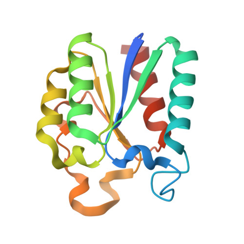
4KIJ

Design and structural analysis of aromatic inhibitors of type II dehydroquinase dehydratase from Mycobacterium tuberculosis - compound 35c [3,4-dihydroxy-5-(3-nitrophenoxy)benzoic acid]


Experimental Data Snapshot

  • Method: X-RAY DIFFRACTION
  • Resolution: 2.80 Å
  • R-Value Free: 0.248 
  • R-Value Work: 0.190 
  • R-Value Observed: 0.193 

wwPDB Validation   3D Report Full Report


Ligand Structure Quality Assessment 


This is version 1.3 of the entry. See complete history


Literature

Design and Structural Analysis of Aromatic Inhibitors of Type II Dehydroquinase from Mycobacterium tuberculosis.

Howard, N.I.Dias, M.V.Peyrot, F.Chen, L.Schmidt, M.F.Blundell, T.L.Abell, C.

(2015) ChemMedChem 10: 116-133

  • DOI: https://doi.org/10.1002/cmdc.201402298
  • Primary Citation of Related Structures:  
    4KI7, 4KIJ, 4KIU, 4KIW

  • PubMed Abstract: 

    3-Dehydroquinase, the third enzyme in the shikimate pathway, is a potential target for drugs against tuberculosis. Whilst a number of potent inhibitors of the Mycobacterium tuberculosis enzyme based on a 3-dehydroquinate core have been identified, they generally show little or no in vivo activity, and were synthetically complex to prepare. This report describes studies to develop tractable and drug-like aromatic analogues of the most potent inhibitors. A range of carbon-carbon linked biaryl analogues were prepared to investigate the effect of hydrogen bond acceptor and donor patterns on inhibition. These exhibited inhibitory activity in the high-micromolar range. The addition of flexible linkers in the compounds led to the identification of more potent 3-nitrobenzylgallate- and 5-aminoisophthalate-based analogues.


  • Organizational Affiliation

    Department of Chemistry, University of Cambridge, Lensfield Road, Cambridge CB2 1EW (UK).


Macromolecules
Find similar proteins by:  (by identity cutoff)  |  3D Structure
Entity ID: 1
MoleculeChains Sequence LengthOrganismDetailsImage
3-dehydroquinate dehydratase153Mycobacterium tuberculosisMutation(s): 0 
Gene Names: aroDaroQMT2612MTCY159.19Rv2537c
EC: 4.2.1.10
UniProt
Find proteins for P9WPX7 (Mycobacterium tuberculosis (strain ATCC 25618 / H37Rv))
Explore P9WPX7 
Go to UniProtKB:  P9WPX7
Entity Groups  
Sequence Clusters30% Identity50% Identity70% Identity90% Identity95% Identity100% Identity
UniProt GroupP9WPX7
Sequence Annotations
Expand
  • Reference Sequence
Small Molecules
Ligands 2 Unique
IDChains Name / Formula / InChI Key2D Diagram3D Interactions
KIJ
Query on KIJ

Download Ideal Coordinates CCD File 
C [auth A]3,4-dihydroxy-5-(3-nitrophenoxy)benzoic acid
C13 H9 N O7
GEHZMCJZQSUKMY-UHFFFAOYSA-N
CL
Query on CL

Download Ideal Coordinates CCD File 
B [auth A]CHLORIDE ION
Cl
VEXZGXHMUGYJMC-UHFFFAOYSA-M
Experimental Data & Validation

Experimental Data

  • Method: X-RAY DIFFRACTION
  • Resolution: 2.80 Å
  • R-Value Free: 0.248 
  • R-Value Work: 0.190 
  • R-Value Observed: 0.193 
  • Space Group: F 2 3
Unit Cell:
Length ( Å )Angle ( ˚ )
a = 126.98α = 90
b = 126.98β = 90
c = 126.98γ = 90
Software Package:
Software NamePurpose
REFMACrefinement
PDB_EXTRACTdata extraction
DNAdata collection
MOSFLMdata reduction
SCALAdata scaling
AMoREphasing

Structure Validation

View Full Validation Report


Ligand Structure Quality Assessment 


Entry History 

Deposition Data

Revision History  (Full details and data files)

  • Version 1.0: 2014-05-14
    Type: Initial release
  • Version 1.1: 2015-01-07
    Changes: Database references
  • Version 1.2: 2018-01-24
    Changes: Structure summary
  • Version 1.3: 2024-02-28
    Changes: Data collection, Database references, Derived calculations