


Crystal Structure of Cofilin Mutant (cof1-157p)
Kish-Trier, E., Haarer, B., Cingolani, G., Amberg, D.C.To be published.
Experimental Data Snapshot
wwPDB Validation 3D Report Full Report

Entity ID: 1 | |||||
|---|---|---|---|---|---|
| Molecule | Chains | Sequence Length | Organism | Details | Image |
| Cofilin | 143 | Saccharomyces cerevisiae S288C | Mutation(s): 1 Gene Names: COF1, YLL050C, L0596 | 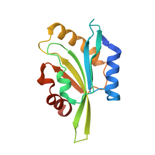 | |
UniProt | |||||
Entity Groups | |||||
| Sequence Clusters | 30% Identity50% Identity70% Identity90% Identity95% Identity100% Identity | ||||
| UniProt Group | Q03048 | ||||
Sequence AnnotationsExpand | |||||
Reference Sequence | |||||
| Length ( Å ) | Angle ( ˚ ) |
|---|---|
| a = 150.023 | α = 90 |
| b = 30.325 | β = 133.01 |
| c = 104.268 | γ = 90 |
| Software Name | Purpose |
|---|---|
| CBASS | data collection |
| PHENIX | model building |
| PHENIX | refinement |
| HKL-2000 | data reduction |
| HKL-2000 | data scaling |
| PHENIX | phasing |


