4KCC

Crystal Structure of the NMDA Receptor GluN1 Ligand Binding Domain Apo State


Experimental Data Snapshot

  • Method: X-RAY DIFFRACTION
  • Resolution: 1.89 Å
  • R-Value Free: 0.206 
  • R-Value Work: 0.172 
  • R-Value Observed: 0.173 

wwPDB Validation   3D Report Full Report


This is version 1.3 of the entry. See complete history


Literature

Conformational Analysis of NMDA Receptor GluN1, GluN2, and GluN3 Ligand-Binding Domains Reveals Subtype-Specific Characteristics.

Yao, Y.Belcher, J.Berger, A.J.Mayer, M.L.Lau, A.Y.

(2013) Structure 21: 1788-1799

  • DOI: https://doi.org/10.1016/j.str.2013.07.011
  • Primary Citation of Related Structures:  
    4KCC, 4KCD

  • PubMed Abstract: 

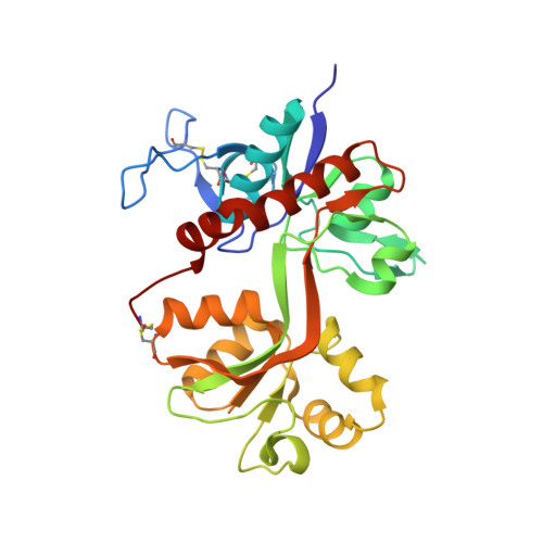
    The NMDA receptor family of glutamate receptor ion channels is formed by obligate heteromeric assemblies of GluN1, GluN2, and GluN3 subunits. GluN1 and GluN3 bind glycine, whereas GluN2 binds glutamate. Crystal structures of the GluN1 and GluN3A ligand-binding domains (LBDs) in their apo states unexpectedly reveal open- and closed-cleft conformations, respectively, with water molecules filling the binding pockets. Computed conformational free energy landscapes for GluN1, GluN2A, and GluN3A LBDs reveal that the apo-state LBDs sample closed-cleft conformations, suggesting that their agonists bind via a conformational selection mechanism. By contrast, free energy landscapes for the AMPA receptor GluA2 LBD suggest binding of glutamate via an induced-fit mechanism. Principal component analysis reveals a rich spectrum of hinge bending, rocking, twisting, and sweeping motions that are different for the GluN1, GluN2A, GluN3A, and GluA2 LBDs. This variation highlights the structural complexity of signaling by glutamate receptor ion channels.


  • Organizational Affiliation

    Laboratory of Cellular and Molecular Neurophysiology, Porter Neuroscience Research Center, National Institute of Child Health and Human Development, Department of Health and Human Services, National Institutes of Health, Bethesda, MD 20892, USA.


Macromolecules
Find similar proteins by:  (by identity cutoff)  |  3D Structure
Entity ID: 1
MoleculeChains Sequence LengthOrganismDetailsImage
Glutamate receptor ionotropic, NMDA 1292Rattus norvegicusMutation(s): 0 
Gene Names: Grin1Nmdar1
UniProt
Find proteins for P35439 (Rattus norvegicus)
Explore P35439 
Go to UniProtKB:  P35439
Entity Groups  
Sequence Clusters30% Identity50% Identity70% Identity90% Identity95% Identity100% Identity
UniProt GroupP35439
Sequence Annotations
Expand
  • Reference Sequence
Small Molecules
Ligands 1 Unique
IDChains Name / Formula / InChI Key2D Diagram3D Interactions
PO4
Query on PO4

Download Ideal Coordinates CCD File 
B [auth A]PHOSPHATE ION
O4 P
NBIIXXVUZAFLBC-UHFFFAOYSA-K
Experimental Data & Validation

Experimental Data

  • Method: X-RAY DIFFRACTION
  • Resolution: 1.89 Å
  • R-Value Free: 0.206 
  • R-Value Work: 0.172 
  • R-Value Observed: 0.173 
  • Space Group: C 1 2 1
Unit Cell:
Length ( Å )Angle ( ˚ )
a = 87.269α = 90
b = 66.359β = 105.21
c = 63.468γ = 90
Software Package:
Software NamePurpose
HKL-2000data collection
PHASERphasing
PHENIXrefinement
HKL-2000data reduction
HKL-2000data scaling

Structure Validation

View Full Validation Report


Entry History 

Deposition Data

Revision History  (Full details and data files)

  • Version 1.0: 2013-07-31
    Type: Initial release
  • Version 1.1: 2013-11-06
    Changes: Database references
  • Version 1.2: 2017-08-02
    Changes: Refinement description, Source and taxonomy
  • Version 1.3: 2023-09-20
    Changes: Data collection, Database references, Derived calculations, Refinement description