Download Mendeley
Novel c-di-GMP recognition modes of the mouse innate immune adaptor protein STING
Chin, K.H., Tu, Z.L., Su, Y.C., Yu, Y.J., Chen, H.C., Lo, Y.C., Chen, C.P., Barber, G.N., Chuah, M.L., Liang, Z.X., Chou, S.H.(2013) Acta Crystallogr D Biol Crystallogr 69: 352-366
- PubMed: 23519410 Search on PubMed
- DOI: https://doi.org/10.1107/S0907444912047269
- Primary Citation Related Structures:
4KBY, 4KC0 - PubMed Abstract:
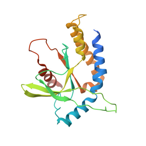
The mammalian ER protein STING (stimulator of interferon genes; also known as MITA, ERIS, MPYS or TMEM173) is an adaptor protein that links the detection of cytosolic dsDNA to the activation of TANK-binding kinase 1 (TBK1) and its downstream transcription factor interferon regulatory factor 3 (IFN3). Recently, STING itself has been found to be the direct receptor of bacterial c-di-GMP, and crystal structures of several human STING C-terminal domain (STING-CTD) dimers in the apo form or in complex with c-di-GMP have been published. Here, a novel set of structures of mouse STING-CTD (mSTING(137-344)) in apo and complex forms determined from crystals obtained under different crystallization conditions are reported. These novel closed-form structures exhibited considerable differences from previously reported open-form human STING-CTD structures. The novel mSTING structures feature extensive interactions between the two monomers, a unique asymmetric c-di-GMP molecule with one guanine base in an unusual syn conformation that is well accommodated in the dimeric interface with many direct specific interactions and two unexpected equivalent secondary peripheral c-di-GMP binding sites. Replacement of the amino acids crucial for specific c-di-GMP binding in mSTING significantly changes the ITC titration profiles and reduces the IFN-β reporter luciferase activity. Taken together, these results reveal a more stable c-di-GMP binding mode of STING proteins that could serve as a template for rational drug design to stimulate interferon production by mammalian cells.
- Agricultural Biotechnology Center, National Chung Hsing University, Taichung 40227, Taiwan.
Organizational Affiliation: