


Crystal structure of the mitochondrial peroxiredoxin from Leishmania braziliensis in the decameric form
Souza, T.A.C.B., Morais, M.A.B., Giuseppe, P.O., Murakami, M.T.To be published.
Experimental Data Snapshot
wwPDB Validation 3D Report Full Report

Entity ID: 1 | |||||
|---|---|---|---|---|---|
| Molecule | Chains | Sequence Length | Organism | Details | Image |
| Peroxidoxin | 192 | Leishmania braziliensis | Mutation(s): 0 Gene Names: LBRM_23_0050 EC: 1.11.1 (PDB Primary Data), 1.11.1.24 (UniProt) | 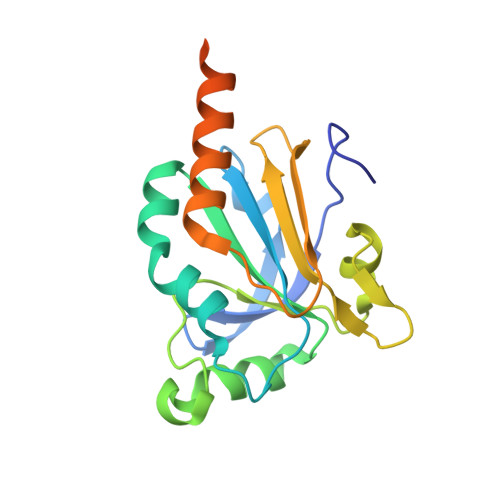 | |
UniProt | |||||
Entity Groups | |||||
| Sequence Clusters | 30% Identity50% Identity70% Identity90% Identity95% Identity100% Identity | ||||
| UniProt Group | A4HCL7 | ||||
Sequence AnnotationsExpand | |||||
Reference Sequence | |||||
| Length ( Å ) | Angle ( ˚ ) |
|---|---|
| a = 90.212 | α = 90 |
| b = 98.901 | β = 90 |
| c = 228.138 | γ = 90 |
| Software Name | Purpose |
|---|---|
| MAR345dtb | data collection |
| MOLREP | phasing |
| PHENIX | refinement |
| XDS | data reduction |
| XDS | data scaling |


