


Crystal Structure of the Ala460Ile mutant of Benzoylformate Decarboxylase from Pseudomonas putida
Brodkin, H.R., Andrews, F.H., Milne, A.C., Petsko, G.A., Ringe, D., McLeish, M.J.To be published.
Experimental Data Snapshot
Starting Model: experimental
View more details
Entity ID: 1 | |||||
|---|---|---|---|---|---|
| Molecule | Chains | Sequence Length | Organism | Details | Image |
| Benzoylformate decarboxylase | 530 | Pseudomonas putida | Mutation(s): 1 Gene Names: mdlC EC: 4.1.1.7 | 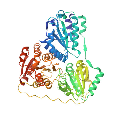 | |
UniProt | |||||
Entity Groups | |||||
| Sequence Clusters | 30% Identity50% Identity70% Identity90% Identity95% Identity100% Identity | ||||
| UniProt Group | P20906 | ||||
Sequence AnnotationsExpand | |||||
Reference Sequence | |||||
| Ligands 3 Unique | |||||
|---|---|---|---|---|---|
| ID | Chains | Name / Formula / InChI Key | 2D Diagram | 3D Interactions | |
| TZD Download:Ideal Coordinates CCD File | G [auth A], J [auth B], L [auth C], O [auth D] | 2-{3-[(4-AMINO-2-METHYLPYRIMIDIN-5-YL)METHYL]-4-METHYL-2-OXO-2,3-DIHYDRO-1,3-THIAZOL-5-YL}ETHYL TRIHYDROGEN
DIPHOSPHATE C12 H18 N4 O8 P2 S ZGJUYGIRPQSCFA-UHFFFAOYSA-N |  | ||
| GOL Download:Ideal Coordinates CCD File | M [auth C] | GLYCEROL C3 H8 O3 PEDCQBHIVMGVHV-UHFFFAOYSA-N |  | ||
| MG Download:Ideal Coordinates CCD File | E [auth A] F [auth A] H [auth B] I [auth B] K [auth C] | MAGNESIUM ION Mg JLVVSXFLKOJNIY-UHFFFAOYSA-N |  | ||
| Length ( Å ) | Angle ( ˚ ) |
|---|---|
| a = 70.637 | α = 90 |
| b = 163.499 | β = 90 |
| c = 175.502 | γ = 90 |
| Software Name | Purpose |
|---|---|
| BlueIce-Epics | data collection |
| PHENIX | model building |
| PHENIX | refinement |
| HKL-2000 | data reduction |
| HKL-2000 | data scaling |
| PHENIX | phasing |


