4JWI

Crystal structure of the substrate binding domain of E.coli DnaK in complex with sheep Bac7(35-43)


Experimental Data Snapshot

  • Method: X-RAY DIFFRACTION
  • Resolution: 1.90 Å
  • R-Value Free: 0.229 
  • R-Value Work: 0.201 
  • R-Value Observed: 0.202 

wwPDB Validation   3D Report Full Report


This is version 1.3 of the entry. See complete history


Literature

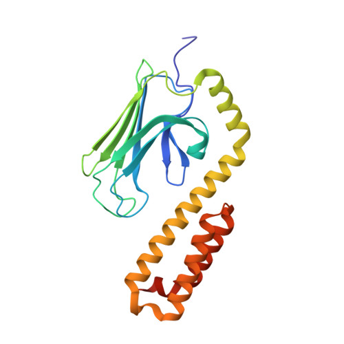
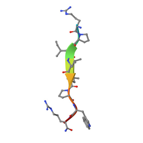
Structural Identification of DnaK Binding Sites within Bovine and Sheep Bactenecin Bac7.

Zahn, M.Kieslich, B.Berthold, N.Knappe, D.Hoffmann, R.Strater, N.

(2014) Protein Pept Lett 21: 407-412

  • DOI: https://doi.org/10.2174/09298665113206660111
  • Primary Citation of Related Structures:  
    4JWC, 4JWD, 4JWE, 4JWI

  • PubMed Abstract: 

    Bacterial resistance against common antibiotics is an increasing health problem. New pharmaceuticals for the treatment of infections caused by resistant pathogens are needed. Small proline-rich antimicrobial peptides (PrAMPs) from insects are known to bind intracellularly to the conventional substrate binding cleft of the E. coli Hsp70 chaperone DnaK. Furthermore, bactenecins from mammals, members of the cathelicidin family, also contain potential DnaK binding sites. Crystal structures of bovine and sheep Bac7 in complex with the DnaK substrate binding domain show that the peptides bind in the forward binding mode with a leucine positioned in the central hydrophobic pocket. In most structures, proline and arginine residues preceding leucine occupy the hydrophobic DnaK binding sites -1 and -2. Within bovine Bac7, four potential DnaK binding sites were identified.


  • Organizational Affiliation

    Institute of Bioanalytical Chemistry, Center for Biotechnology and Biomedicine, University of Leipzig, Deutscher Platz 5, 04103 Leipzig, Germany. strater@bbz.uni-leipzig.de.


Macromolecules
Find similar proteins by:  (by identity cutoff)  |  3D Structure
Entity ID: 1
MoleculeChains Sequence LengthOrganismDetailsImage
Chaperone protein DnaK
A, B
219Escherichia coli K-12Mutation(s): 0 
Gene Names: dnaKgroPgrpFseg
UniProt
Find proteins for P0A6Y8 (Escherichia coli (strain K12))
Explore P0A6Y8 
Go to UniProtKB:  P0A6Y8
Entity Groups  
Sequence Clusters30% Identity50% Identity70% Identity90% Identity95% Identity100% Identity
UniProt GroupP0A6Y8
Sequence Annotations
Expand
  • Reference Sequence

Find similar proteins by:  Sequence   |   3D Structure  

Entity ID: 2
MoleculeChains Sequence LengthOrganismDetailsImage
Cathelicidin-3
C, D
10Ovis ariesMutation(s): 1 
UniProt
Find proteins for P50415 (Ovis aries)
Explore P50415 
Go to UniProtKB:  P50415
Entity Groups  
Sequence Clusters30% Identity50% Identity70% Identity90% Identity95% Identity100% Identity
UniProt GroupP50415
Sequence Annotations
Expand
  • Reference Sequence
Experimental Data & Validation

Experimental Data

  • Method: X-RAY DIFFRACTION
  • Resolution: 1.90 Å
  • R-Value Free: 0.229 
  • R-Value Work: 0.201 
  • R-Value Observed: 0.202 
  • Space Group: P 21 21 2
Unit Cell:
Length ( Å )Angle ( ˚ )
a = 77.914α = 90
b = 161.159β = 90
c = 45.114γ = 90
Software Package:
Software NamePurpose
MAR345data collection
BUSTERrefinement
XDSdata reduction
SCALAdata scaling

Structure Validation

View Full Validation Report


Entry History 

Deposition Data

Revision History  (Full details and data files)

  • Version 1.0: 2013-11-13
    Type: Initial release
  • Version 1.1: 2014-03-26
    Changes: Database references
  • Version 1.2: 2017-11-15
    Changes: Refinement description
  • Version 1.3: 2023-09-20
    Changes: Data collection, Database references, Derived calculations, Refinement description