4JUP

Dimeric structure of CARMA1 CARD


Experimental Data Snapshot

  • Method: X-RAY DIFFRACTION
  • Resolution: 3.20 Å
  • R-Value Free: 0.388 
  • R-Value Work: 0.259 
  • R-Value Observed: 0.265 

wwPDB Validation   3D Report Full Report


This is version 1.1 of the entry. See complete history


Literature

Novel Disulfide Bond-Mediated Dimerization of the CARD Domain Was Revealed by the Crystal Structure of CARMA1 CARD

Jang, T.H.Park, J.H.Park, H.H.

(2013) PLoS One 8: e79778-e79778

  • DOI: https://doi.org/10.1371/journal.pone.0079778
  • Primary Citation of Related Structures:  
    4JUP

  • PubMed Abstract: 

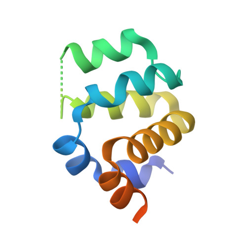
    CARMA1, BCL10 and MALT1 form a large molecular complex known as the CARMA1 signalosome during lymphocyte activation. Lymphocyte activation via the CARMA1 signalosome is critical to immune response and linked to many immune diseases. Despite the important role of the CARMA1 signalosome during lymphocyte activation and proliferation, limited structural information is available. Here, we report the dimeric structure of CARMA1 CARD at a resolution of 3.2 Å. Interestingly, although CARMA1 CARD has a canonical six helical-bundles structural fold similar to other CARDs, CARMA1 CARD shows the first homo-dimeric structure of CARD formed by a disulfide bond and reveals a possible biologically important homo-dimerization mechanism.


  • Organizational Affiliation

    School of Biotechnology and Graduate School of Biochemistry at Yeungnam University, Gyeongsan, South Korea.


Macromolecules
Find similar proteins by:  (by identity cutoff)  |  3D Structure
Entity ID: 1
MoleculeChains Sequence LengthOrganismDetailsImage
Caspase recruitment domain-containing protein 11
A, B
104Homo sapiensMutation(s): 1 
Gene Names: CARD11CARMA1
UniProt & NIH Common Fund Data Resources
Find proteins for Q9BXL7 (Homo sapiens)
Explore Q9BXL7 
Go to UniProtKB:  Q9BXL7
PHAROS:  Q9BXL7
GTEx:  ENSG00000198286 
Entity Groups  
Sequence Clusters30% Identity50% Identity70% Identity90% Identity95% Identity100% Identity
UniProt GroupQ9BXL7
Sequence Annotations
Expand
  • Reference Sequence
Experimental Data & Validation

Experimental Data

  • Method: X-RAY DIFFRACTION
  • Resolution: 3.20 Å
  • R-Value Free: 0.388 
  • R-Value Work: 0.259 
  • R-Value Observed: 0.265 
  • Space Group: P 21 21 21
Unit Cell:
Length ( Å )Angle ( ˚ )
a = 45.887α = 90
b = 53.863β = 90
c = 92.802γ = 90
Software Package:
Software NamePurpose
HKL-2000data collection
PHASESphasing
REFMACrefinement
HKL-2000data reduction
HKL-2000data scaling

Structure Validation

View Full Validation Report


Entry History 

Deposition Data

  • Released Date: 2014-02-05 
  • Deposition Author(s): Park, H.H.

Revision History  (Full details and data files)

  • Version 1.0: 2014-02-05
    Type: Initial release
  • Version 1.1: 2018-02-07
    Changes: Experimental preparation